

Overview
The knee menisci are two crescenteric fibrocartilaginous pads with a triangular cross-section that sits within medial and lateral aspects of the knee joint, deepening the tibifemoral articulation. It’s primary roles are shock absorption, improving stability and load transmission.
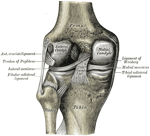
Surfaces
- The superior surfaces of menisici are concave
- The inferior surface is flat to accomodate the tibial plateau
Structure
Medial Meniscus
- Larger than the lateral meniscus and broader posteriorly than it is anteriorly
- The anterior horn is attached to the tibia plataeu near the intercondylar fossa anterior ot the anterior cruciate ligament.
- The posterior horn is attach anterior to the posterior cruciate ligament.
- Average width of 9 to 10mm, average thickness of 3 to 5mm
Lateral Meniscus
- Lateral meniscus is more circular with the horns closer together
- The anterior horn is attached immediately lateral to the tibial attachment of the anterior cruciate ligament
- The posterior horn is attached anterior to medial meniscus and posterior cruciate ligament
- Covers a larger portion of the articular surface
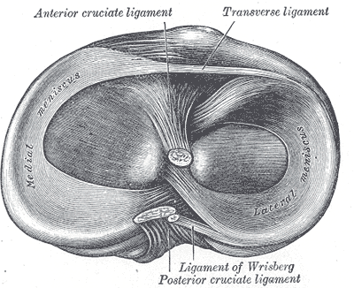
Attachments
Medial meniscus
- The anterior horn attaches immediately anterior to the tibial attachment of the anterior cruciate ligament (ACL) in the intercondylar area
- The medial aspect attached to the deep (third) layer of the medial collateral ligament
- The posterior horn is attached to the posterior intercondylar area of the tibial plateau, between PCL insertion (posteriorly) and posterior root attachment of lateral meniscus (anteriorly)
Lateral meniscus
- The anterior horn of the lateral meniscus attaches immediately lateral to the tibial attachment of the ACL on the intercondylar area
- No attachment to the lateral collateral ligament
- Attached to joint capsule except anterior horn and posterior most portion of posterior horn; this is due to the passage of the intra-articular portion of the popliteus tendon
- Posterior horn of the lateral meniscus attaches to the posterior intercondylar area of the tibial plateau anterior to the medial meniscus and posterior to ACL
Meniscofemoral ligament
- Arises from the posterior horn of the lateral meniscus and attaches to the lateral aspect of the medial femoral condyle. It splits into two bands at the posterior crucial ligament, named in accordance to its relation with it:
- Anterior meniscofemoral ligament (of Humphrey) – anterior to the PCL
- Posterior meniscofemoral ligament (of Wrisberg) – posterior to the PCL
Oblique menisco-meniscal ligament
Transverse meniscal (or genual) ligament (of Winslow)
- Attaches the anterior horns to each other
Arterial Supply
The menisci are vascularized only via the periphery and the root attachments and therefore the inner portions are avascular. This has important implications on the healing and surgical management of meniscal tears.
- Red zone (outer one-third) – supply from the peripheral meniscal plexus, formed by the medial, lateral and middle genicular arteries
- White zone (inner two-thirds) – no vascular supply
- Red-white zone (transition between outer third and inner two-thirds) – gradual loss of perfusion
Innervation
Posterior articular branch of the tibial nerve and terminal obturator and femoral nerve branches
Variants
- Discoid meniscus – body that is too wide, prone to cystic degeneration with subsequent tear (usually affecting the lateral meniscus)
- Congenital hypoplasia or absence
- Speckled anterior horn of lateral meniscus
- Anomalous insertion of the medial meniscus (AIMM) (2%)
- Anterior root onto the ACL or intercondylar notch
- Anterior margin of the tibia in the midline
- No attachment: stabilized by the transverse meniscal ligament 5
- Meniscal fluonce
- Meniscal ossicle