

Overview
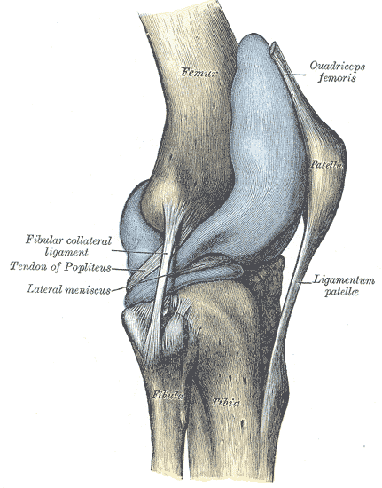
The patella (kneecap) is a thick, flat, triangular-shaped sesamoid bone (largest sesamoid in the human body) located anterior to the knee joint within tendon of the quadriceps femoris muscle. It has two main functions:
- Leg extension – Improve the effective extension capacity of the quadriceps muscle by increasing the moment arm of the patellar ligament
- Protection – Protects the quadriceps tendon from frictional forces by minimizing tendon contact with the femur and acts as a bony shield again physical trauma for deeper structures in the knee joint
Structure & Shape
The patella is a uniformly dense trabecular bone with a thin compact lamina covering it. The Wiberg classification system describes the shape based on the asymmetry between the medial and lateral facets on axial view of the patella.
- Wiberg Type 1/a (see in 10% of population)
- Roughly symmetrical facets
- Concave facets
- Equally sized facets
- Wiberg Type 2/b
- Slightly smaller size of the medial facet
- Concave aspect of the lateral facet
- Wiberg Type 3/c
- Markedly smaller size of the medial facet
- More vertical orientation of the medial facet
Surfaces & Articulations
Anterior surface
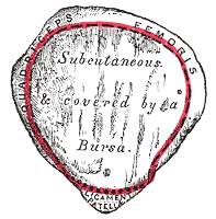
Convex in all directions and perforated by small apertures for the passage of nutrient vessels. The surface is marked by numerous rough longitudinal striae and separated from the integument by a bursa.
- Base – thickened, roughed border which forms the superior aspect of the patella and provides attachment for the quadriceps tendon
- Apex – points inferiorly, connected to the tibial tuberosity via the patella ligament
Posterior surface
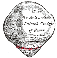
The superior two thirds of the posterior articular surface of patella articulates with the anterior surface of the femoral condyles to form the patellofemoral joint and is covered with articular cartilage. By comparison to the anterior surface, it is smooth and divided into two facets by a vertical ridge which corresponds to the groove on the patelalr surface of the femur.
- Medial facet – articulates with the medial condyle of the femur
- Odd facet – exists along the extreme medial border of the medial facet
- Lateral facet – broader surface area than the medial facet, articulates with the lateral condyle of the femur
- Note: interestingly, it is the medial femoral condyler which is larger and projects more anteriorly than the lateral condyle

Arterial Supply
A peripatella anastomotic arterial ring supplies the patella. It is contributed by genicular arteries, branches of the popliteal artery:
- Superior medial and lateral genicular artery
- Inferior medial and lateral genicular artery
- Descending and anterior genicular artery
Venous Drainage
Draining veins follow the genicular artery and are accompanied by a complementary system of veins running deep to the articular cartilage forming a subarticular drainage network
Innervation
- Branches of nerves to vastus medialis and vastus lateralis
Attachments
The superior third of the patella acts as the attachment point for the tendinous fibres of the rectus femoris and vastus intermedius of the qaudriceps.
- Quadriceps tendon (superiorly) – continous with the patellar tendon (considered the same structure)
- Patellar tendon – attaches to the tibial tubercle inferiorly
- Medial and lateral patellar retinaculum – condensations of fascia attach the patella margins to surrounding fascia.
- Medial patellar retinaculum – attaches to the vastus medialis / sartorius fascia
- Lateral patellar retinaculum – attaches to the fascia of vastus lateralis and the iliotibial band
- The medial patellofemoral ligament (MPFL) originates near adductor tubercle of the femur and inserts into the superomedial aspect of the patella. Its function is to prevent lateral patellar dislocation during knee extension.
Development
The bone originates from multiple ossification centres that develop from the ages of three to six, which rapidly coalesce

Relations
- Superior: quadriceps tendon, suprapatellar bursa
- Inferior: patellar tendon, infrapatellar bursa, infrapatellar (Hoffa’s) fat pad
- Lateral: lateral patellar retinaculum
- Medial: medial patellar retinaculum
- Posterior: knee joint, femur
- Anterior: prepatellar bursa
Anatomical Variants
- Absent patella
- Bipartite patella
- Multipartite patella
- Emargination of the proximal lateral edge
- Hypoplastic
- Dorsal defect of the patella
- Variation in shape (shaped classified by the Wiberg classification)