

Pelvic Floor |
|---|
| CATEGORY 1’20 | ’19 | ’18 | ’10 |
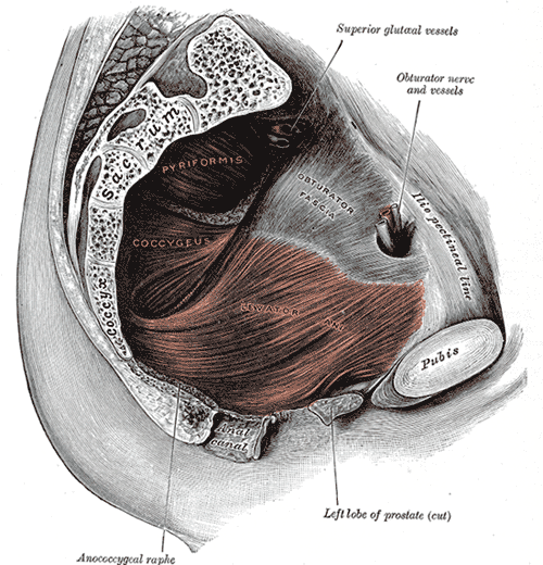
The pelvic floor (or diaphragm) is a bowl-shaped muscular part of the pelvic floor, composed of muscle fibres of the levator ani (a muscle group), coccygeus muscle and connective tissue.
Levator Ani |
|---|
| CATEGORY 1’20 | ’19 | ’18 | ’10 |
The levator ani is a broad, thin muscle group situated on either side of the pelvis. It is formed by three paired muscles; pubococcygeus, iliococcygeus and puborectalis. It is the largest component of the pelvic floor.

Origin and Insertion
- Anterior: The posterior surface of the superior pubic ramus, lateral to the symphysis
- Laterally: Tendinous arch – thickened band in the fascia covering obturator internus muscle
- Posteriorly: The inner surface of the spine of the ischium
Puborectalis
- Originates from both sides of the body of the pubis, and passes posteriorly to encircle the rectum and form a U-shaped sling around the anorectal junction (thus no insertion point as such).
- Tonic contraction bends the canal anteriorly and forms a 90-degree anorectal angle at the anorectal junction to maintain faecal continence
Pubococcygeus
- Main component of levator ani
- Originates from the body of pubis, lateral to the origin of the puborectalis muscle
- The fibres course around margin of urogenital hiatus posteriorly to attach to the tendinous centre of the perineum, anococcygeal body and coccyx
- Supports pelvic organs
Iliococcygeus
- Originates from the inner surface of the ischial spine and along tendinous arch (a thickened band of fascia covering the inner aspect of obturator internus muscle)
- Posteriorly attaches to iliococcygeus of the opposite side in the midline to form anococcygeal raphe extending from anal aperture to coccyx
- Supports pelvic organs
Blood supply
- Inferior gluteal artery – branch of internal iliac artery (anterior division)
- Inferior vesical – branch of internal iliac artery (anterior division)
- Pudendal
Innervation
- Innervated by nerve to levator ani, branch from ventral ramus of S4
- Also contributions from inferior rectal branch of pudendal nerve (S2-4)
Relations
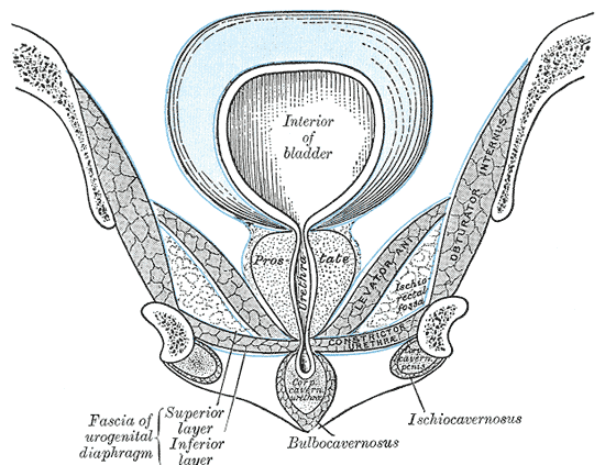
- Inferiorly: perineum (anus), ischiorectal fossa, urogenital diaphragm
- Medially: prostate, urethra
- Superiorly: bladder, uterus (female)
- Anteriorly: pubic rami
Apertures
- Urogenital hiatus – U-shaped defect anteriorly in levator ani, allowing passage of urethra (and vagina in females)
- Rectal hiatus – centrally positioned defect allowing passage of anal canal
o Sphincter urethrovaginalis (in women)
o Compressor urethrae (in women)
Variants
- Sacral spinal nerves (S3, S4) innervate the muscles directly as well (~70%).
- Inferior rectal nerve innervates the levator ani muscles independently of the pudendal nerve (~40%)
Coccygeus |
|---|
| CATEGORY 1’20 | ’19 | ’18 | ’10 |
The coccygeus is a triangular musculotendinous sheet overlying the pelvic surface of sacrospinous ligament. It is a smaller, most posterior pelvic floor component.
Origin and Insertion
- Apex attached to tip of ischial spine
- Base attached to lateral margin of coccyx and fifth sacral segment
- Innervated by anterior rami of S2-4
Relations
- Anteriorly: levator ani
- Posteriorly: sacrospinous ligament
Innervation
- Pudendal nerve
- The pudendal nerve runs between the coccygeus muscle and the piriformis muscle, superficial to the coccygeus muscle
Blood supply
- Inferior gluteal artery
- Inferior vesical
- Pudendal