

Overview
The pineal gland or body (epiphysis) is a small (approximately 6mm), pea-sized neuroendocrine gland that comprises part of the epithalamus (component of the diencephalon). It is located in the midline of the brain between the two superior colliculi. It is attached by a stalk to the posterior wall of the third ventricle, above the quadrigeminal plate. Its main function is to secrete melatonin, which regulates the circadian rhythm of the body.
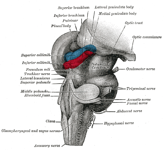
Structure
Pineal stalk
- Divided into the superior and inferior laminae, which contains the habenular and posterior commisure respectively
Pineal recess
- The space between the laminae
- Filled with an extension of the cavity of the third ventricle
Corpora areneca
- ‘Brain sand’ – deposits of calcium, phosphates and carbonates forming multilaminar corpuscles
Arterial Supply
Posterior choroidal arteries – branches that arise from the posterior cerebral artery
Venous Drainage
Internal cerebral veins
Relations
Superior
- Splenium of the corpus callosum
- Internal cerebral veins
- vein of Galen (posteriorly)
- stria medullaris, splenium of the corpus callosu
- Velum interpositum
Inferior
- Superior colliculi of the midbrain
Anteriorly
- Third ventricle (pineal recess)
- Anterosuperiorly: habenular
- Anteroinferiorly: posterior commissure
Posterior
- Posteroinferiorly: superior cerebellar cistern