

April 2013
Question 1
- The ICRP system of radiation protection is founded on the twin tenets of Justification and Optimization backed up by the application of dose limits.
- In the specific context of the medical use of ionizing radiation discuss what is meant by Justification and Optimization and give examples of how these principles might be applied in practice (7 marks)
- Dose limits do not apply to patient exposures but increasingly Diagnostic Reference Levels (DRLs) do. What are DRLs, and how can they be used to optimize imaging procedures in diagnostic radiology? (5 marks)
- A pregnant woman undergoes the following examinations at various stages of her pregnancy as outlined below. Provide an estimate of the magnitude of any foetal radiation doses involved, discuss any possible risks to the foetus following each procedure and indicate what you might say to the concerned patient to allay any fears she may have. (8 marks)
- CT of the abdomen and pelvis with and without contrast at 7 weeks
- Mammography at 8 weeks
- Ultrasound of the liver at 10 weeks.
- Ventilation/perfusion lung scan at 14 weeks.
Question 2
- Define and briefly describe each of the following in the context of X-ray interactions in the diagnostic energy range:
- Photoelectric effect (2 marks)
- Compton scatter (2 marks)
- Linear attenuation coefficient (1 mark)
- Mass attenuation coefficient (1 mark)
- Half value layer (1 mark)
- The basic components of a fluoroscopic X-ray system suitable for interventional procedures consists of a generator, X-ray tube and housing including collimator and filtration, patient table, anti-scatter grid, and an image receptor Describe the key design features and function of each of these components and briefly discuss the role of automatic brightness control.(9 marks)
- Briefly describe the technical basis and application of each of the following in the context of DSA image processing:
- Logarithmic subtraction (2 marks)
- Reregistration (pixel shift) (1 mark)
- Roadmapping (1 mark)
Question 3
- The patient dose from a helical CT scan is affected by a number of machine parameters that are controllable by the operator. List five (5) of these parameters and describe how they affect patient dose and why. (5 marks)
- Contrast in an MRI image depends on three (3) parameters intrinsic to the tissue being imaged. Describe these three parameters and explain what operator adjustable factors are required to cause each of them to predominate in a spin echo sequence, giving reasons for your selected factors. In the sequences where relaxation times give the most contrast, what values of the intrinsic parameters will appear brightest? (5 marks)
- Describe the basics of ultrasound pulse-echo imaging. In addition, list the factors that determine the amplitude of the echo received by the transducer. (5 marks)
-
- Collimator design has a significant influence on the spatial resolution and sensitivity of a gamma camera. For a parallel hole collimator explain how collimator hole size & length and septum thickness affects system spatial resolution and sensitivity. (3 marks)
- List six properties that a radionuclide would have to make it suitable for use in single photon imaging. (2 marks)
September 2013
Section 1 – Radiation Biology and Safety
Question 1
- Ionizing radiation may cause stochastic effects or deterministic effects (tissue reactions) in human species. Briefly explain what is meant by these outcomes and provide two examples of each. (5 marks)
- The ICRP has developed a system of dose limits for the protection of individuals and populations. What are the dose limits for stochastic effects for occupational exposed individuals and members of the public, respectively? Why is there a difference between the limits for occupationally exposed workers and members of the public? What is the dose limit that applies once a female employee becomes pregnant? (5 marks)
Question 2
An obese female patient of child bearing age presents with clinical indications of renal colic. A CT abdominal examination (KUB) is suggested and is undertaken on the basis that she is not pregnant. On viewing the reconstructed images it becomes apparent that she was pregnant. She subsequently undergoes an ultrasound examination which establishes that the fetal age at the time of the CT examination was 14 weeks. What can you tell the patient about any possible concerns for the health of the fetus? Your response should start with an estimate of the fetal dose. (10 marks)
Question 3
A fixed under-table X-ray system is used for general purpose fluoroscopy on adult patients. The X-ray unit has automatic brightness control (ABC) with a range of dose rate options, including pulsed fluoroscopy and an image intensifier with variable field size. Discuss practical procedural measures that you might implement to ensure that the patient dose is minimised without compromising the diagnostic information obtained. Supplement your answer with reasons for your actions. (10 marks)
Section 2 – Basic Physics & Technology including Mammography, Fluoroscopy & DSA
Question 1
- Distinguish between the terms contrast and spatial resolution as applied in diagnostic radiography. (4 marks)
- X-ray tubes (XRTs) typically consist of an anode and cathode mounted inside an evacuated envelope. Tubes used in general radiography and mammography are similar in this regard, but they also differ in a number of important design features. Write a detailed account about two of these which impact directly on the contrast and spatial resolution of radiographs. (6 marks)
Question 2
- Explain why scattered radiation is generally of concern in X-ray imaging. (3 marks)
- Describe the influence of three factors which have an impact on the generation of scatter in radiographs.(3 marks)
- Briefly explain how an anti-scatter grid works. (3 marks)
- Computed radiographs of the skull are acquired at 75 kV with and without a grid. The sensitivity number (S-number) for each radiograph was ~100. The exposures with and without the grid were 20 mAs and 4 mAs, respectively. Calculate the grid’s Bucky Factor. (1 mark)
Question 3
- In the context of chest radiography, briefly describe the design and operation of the Automatic Exposure Control (AEC) device? (5 marks)
- In the context of fluoroscopy of the digestive tract, briefly describe the function and operation of the Automatic Brightness Control (ABC) system? (5 marks)
Section 3 – CT, MRI, US & Nuclear Medicine
Question 1
- Most clinical CT scanners today are multi-detector CT (MDCT) units. Briefly describe the MDCT scanner geometry and the function of three key physical parameters under operator control for a CT helical acquisition. (4 marks)
- What is meant by partial volume artefact? Describe how this effect can be mitigated and what are the possible effects of this on image quality and patient dose? (6 marks)
Question 2
- The Larmor precession is a fundamental process in nuclear resonance. Explain the concept of Larmor precession, including Larmor frequency and explain its significance in MRI imaging. (4 marks)
- Tissues have two relaxation parameters, intrinsic to the tissue being imaged, that can be used to provide contrast in MRI images. Name the two relaxation parameters and describe one of the parameters in detail. (3 marks)
- Safety in MRI is an important consideration in clinical practice. Identify three safety issues (patient and environmental) or contra-indications in the use of MRI. (3 marks)
Question 3
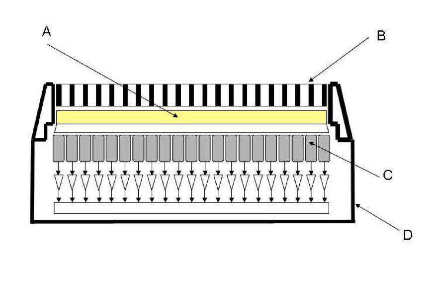
- Gamma cameras are the basic imaging device for much of nuclear medicine imaging. The diagram depicts a schematic of the major components of a gamma camera detector head. For each of the components labelled A to D in the diagram, name the component and briefly describe its function in the detector head’s operation. (6 marks)
- In Positron Emission Tomography imaging there are two fundamental physical factors that limit the ultimate spatial resolution achievable by any PET camera. State each of these factors and briefly explain how each one limits the spatial resolution achievable. (4 marks)