

Cerebellum
The cerebellum is a major structure of the hindbrain responsible for coordinating voluntary movements, balance, posture, and motor learning. Although it constitutes only about 10% of the brain’s volume, it contains more than 50% of the brain’s neurons. The cerebellum communicates with the cerebral cortex, brainstem, and spinal cord to fine-tune motor activity and ensure smooth execution of movement patterns.
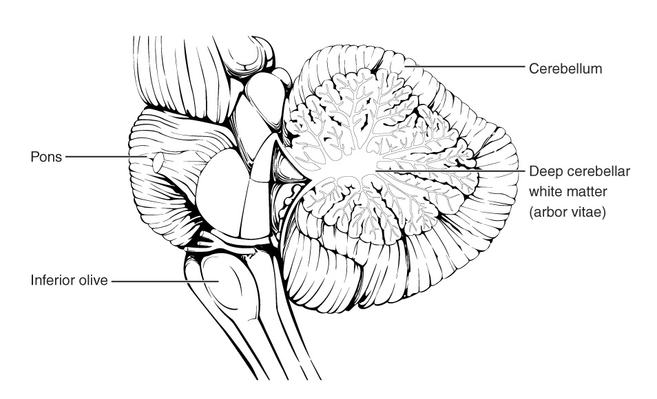
The cerebellum is located in the posterior cranial fossa, beneath the occipital lobes of the cerebrum and dorsal to the brainstem. It is separated from the cerebral hemispheres by the tentorium cerebelli, a dural fold that helps compartmentalize the supratentorial and infratentorial structures.
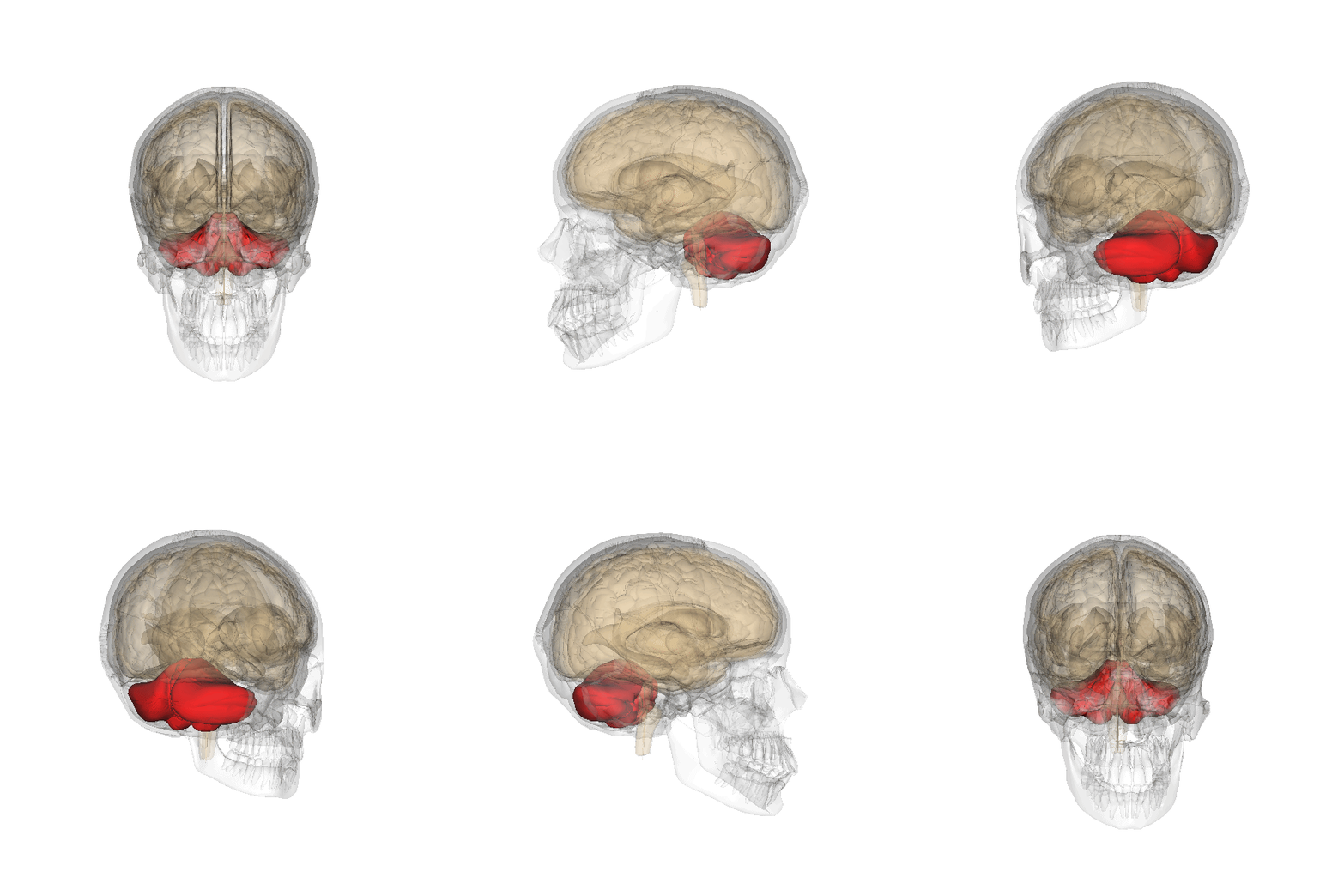
Relational Anatomy
- Anterior: Brainstem (pons and medulla oblongata)
- Posterior: Occipital bone and cisterna magna
- Superior: Tentorium cerebelli and occipital lobes
- Inferior: Foramen magnum and spinal cord
- Lateral: Cerebellopontine angle (containing the cranial nerves VII and VIII)
Development
The cerebellum develops from the rhombic lip of the metencephalon, part of the embryonic hindbrain. By the third trimester, the cerebellum has fully formed its lobes and begins refining its neural circuits postnatally. Cerebellar growth continues throughout childhood, with the most rapid development occurring during the perinatal period [3].
5. Function
The cerebellum is primarily responsible for:
- Motor coordination: Regulating the force, timing, and precision of voluntary movements
- Balance and posture: Integrating sensory input to maintain equilibrium
- Motor learning: Adapting movements through practice, such as in sports or musical training
- Cognitive roles: Contributing to language, attention, and problem-solving
Structure
Gross Anatomy
The cerebellum consists of two hemispheres connected by the vermis (a midline structure). It is divided into three lobes:
- Anterior lobe (involved in proprioception and muscle tone)
- Posterior lobe (largest lobe, associated with voluntary motor coordination)
- Flocculonodular lobe (important for balance and eye movement control)
Each hemisphere contains three functional zones:
- Vermis (midline zone): Controls axial and proximal limb muscles
- Intermediate zone (paravermal region): Controls distal limb coordination
- Lateral zone: Involved in motor planning and cognitive functions
6.3 Radiological Anatomy
Best Imaging Modality:
- MRI is the modality of choice for cerebellar assessment due to its superior soft-tissue resolution.
- CT is useful for detecting acute hemorrhage, hydrocephalus, or mass effect.
Preferred Planes and Sequences:
- Axial and coronal planes provide the best anatomical overview.
- T2-weighted MRI highlights cerebellar white matter and CSF-filled spaces.
- FLAIR sequences help detect demyelination and gliosis.
- DWI (diffusion-weighted imaging) is essential for detecting acute infarcts.
Normal Cerebellar Dimensions:
- Average transverse diameter: 8–10 cm
- Average height: 4–5 cm
7. Arterial Supply
The cerebellum is supplied by three pairs of arteries originating from the vertebrobasilar system:
- Superior cerebellar artery (SCA) – arises from the basilar artery
- Anterior inferior cerebellar artery (AICA) – arises from the basilar artery
- Posterior inferior cerebellar artery (PICA) – arises from the vertebral artery
8. Venous Drainage
Venous drainage is through the superior and inferior cerebellar veins, which drain into the transverse sinus, sigmoid sinus, and straight sinus.
9. Lymphatic Drainage
Though the brain lacks a traditional lymphatic system, the cerebellum drains interstitial fluid through the glymphatic system, which connects to the deep cervical lymph nodes.
10. Innervation
The cerebellum does not receive direct motor innervation but interacts with the brainstem via:
- Inferior cerebellar peduncle (ICP): Connects to the medulla and carries sensory input from the spinal cord
- Middle cerebellar peduncle (MCP): Connects to the pons and transmits corticopontocerebellar fibers
- Superior cerebellar peduncle (SCP): Connects to the midbrain and carries cerebellar output to the thalamus and cortex
11. Normal Variants
- Mild vermian hypoplasia – can be incidental but may also be associated with developmental syndromes.
- Mega cisterna magna – an enlarged posterior fossa CSF space that can mimic Dandy-Walker malformation.
- Cerebellar tonsillar ectopia – a mild form of Chiari malformation if the tonsils extend ≥5 mm below the foramen magnum.
12. Common Pathologies
- Cerebellar infarcts (PICA, SCA, AICA territory strokes)
- Cerebellar atrophy (due to neurodegenerative disorders or chronic alcohol use)
- Cerebellar tumors (e.g., medulloblastoma in children, hemangioblastoma in adults)
References
- Schmahmann JD. The cerebellum and cognition. Neuroscience & Biobehavioral Reviews. 2019;105:70-85.
- Voogd J, Glickstein M. The anatomy of the cerebellum. Trends in Neurosciences. 1998;21(9):370-375.
- Wang VY, Zoghbi HY. Genetic regulation of cerebellar development. Nature Reviews Neuroscience. 2001;2(7):484-491.