Meniscus
The menisci are critical structures within the knee joint that play an essential role in load distribution, shock absorption, and joint stability. These fibrocartilaginous, crescent-shaped pads sit between the femur and tibia, with each knee containing two menisci: the medial and lateral.

Gross Anatomy of the Menisci
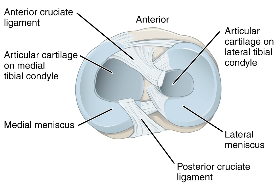
The menisci are located on the tibial plateau, between the femoral condyles and tibial surface. They are thicker at the periphery and taper towards the centre. The medial meniscus is C-shaped, while the lateral meniscus is nearly circular.
Medial Meniscus
- Larger and more fixed than the lateral meniscus.
- Attached firmly to the medial collateral ligament (MCL) and the tibial plateau.
- More prone to injury due to its limited mobility.
Lateral Meniscus
- Smaller and more mobile.
- Has no attachment to the lateral collateral ligament (LCL), allowing it greater freedom of movement.
- Less commonly injured due to its mobility.
Function
The primary functions of the menisci include:
- Load Transmission: The menisci help transmit weight across the knee joint. They bear about 50-70% of the load during daily activities, such as walking.
- Shock Absorption: Acting as cushions, they distribute forces to reduce the impact on articular cartilage.
- Joint Stability: They deepen the articular surface, enhancing the stability of the femoral condyles on the tibial plateau.
- Lubrication: The menisci help spread synovial fluid across the joint, ensuring smooth motion.
Vascular Supply
The blood supply to the menisci is crucial to understanding their healing potential. The menisci are divided into three zones based on vascular supply:
- Red-Red Zone: The peripheral 10-30% of the meniscus, which is richly vascularised, offering the best potential for healing.
- Red-White Zone: The intermediate zone with limited vascularity.
- White-White Zone: The central zone, which is avascular, meaning injuries here are unlikely to heal on their own.
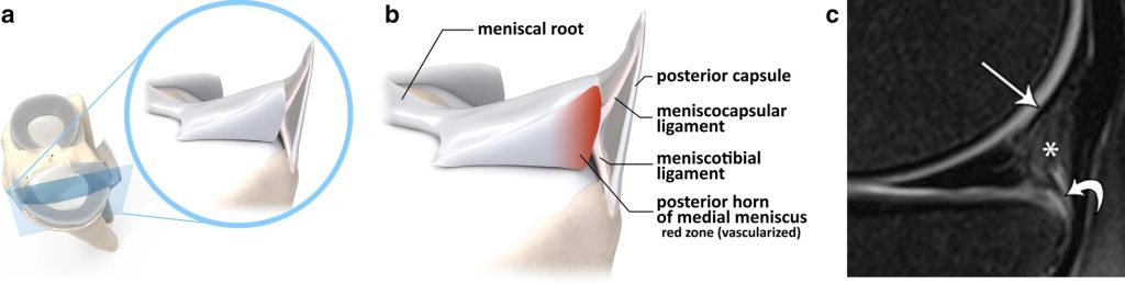
a) Open view of the medial compartment of the knee
b) Magnified view of the posteromedial capsular-meniscal unit
(c) Sagittal PD-weighted fat-suppressed MRI: meniscocapsular ligament (thin arrow), meniscotibial ligament (curved arrow) and posterior capsular attachment (star)
Imaging
MRI is the imaging modality of choice for meniscal assessment. T2-weighted MRI sequences are particularly useful as they provide high contrast between the meniscus (which appears dark) and surrounding joint structures.
- Normal Meniscus Appearance on MRI:
- On T1 and T2-weighted images, the normal meniscus appears as a uniform, low-signal (dark) structure.
- The wedge-shaped appearance in coronal and sagittal sections corresponds to the fibrocartilaginous structure.