

Pituitary Gland
The pituitary gland, often termed the “master gland,” plays a pivotal role in regulating various hormonal functions throughout the body. Despite its small size, its impact on growth, metabolism, and reproductive health is profound.
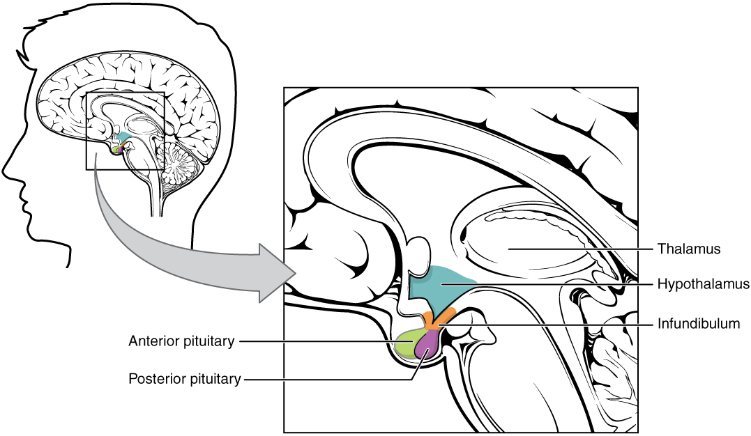
The pituitary gland is located at the base of the brain, seated within the sella turcica, a bony cavity of the sphenoid bone. It lies just inferior to the hypothalamus, to which it is connected by the pituitary stalk (infundibulum). The gland is situated within the middle cranial fossa, surrounded by the cavernous sinuses laterally and positioned below the optic chiasm.
Structure of the Pituitary Gland
The pituitary gland is a bilobed structure, divided into:
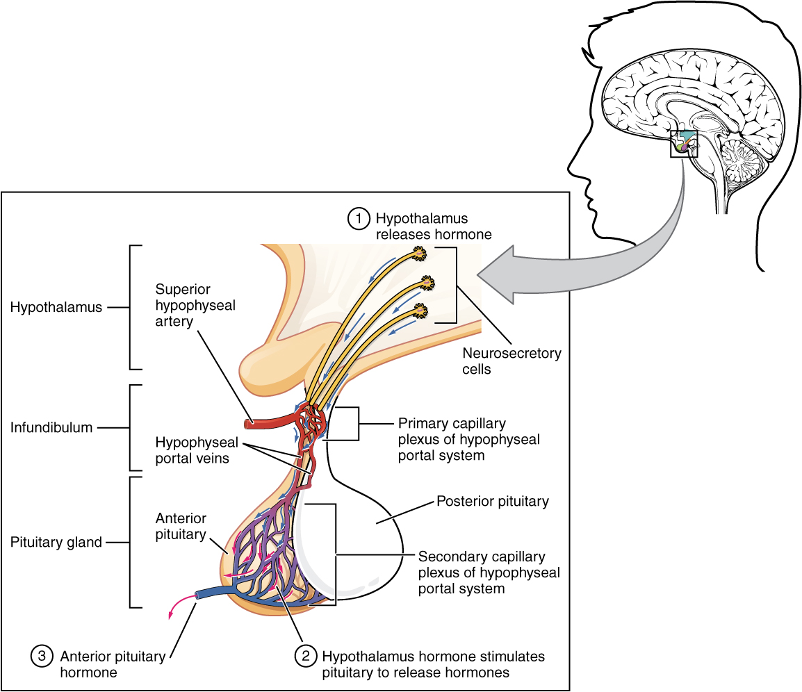
- Anterior Pituitary (Adenohypophysis): Comprising about 80% of the gland, it originates from Rathke’s pouch, an embryonic structure. This part is responsible for hormone production and secretion.
- Posterior Pituitary (Neurohypophysis): Composed mainly of nerve fibers originating from the hypothalamus, it stores and releases hormones produced in the hypothalamus.
Comprising about 80% of the gland, it originates from Rathke’s pouch, an embryonic structure. This part is responsible for hormone production and secretion.
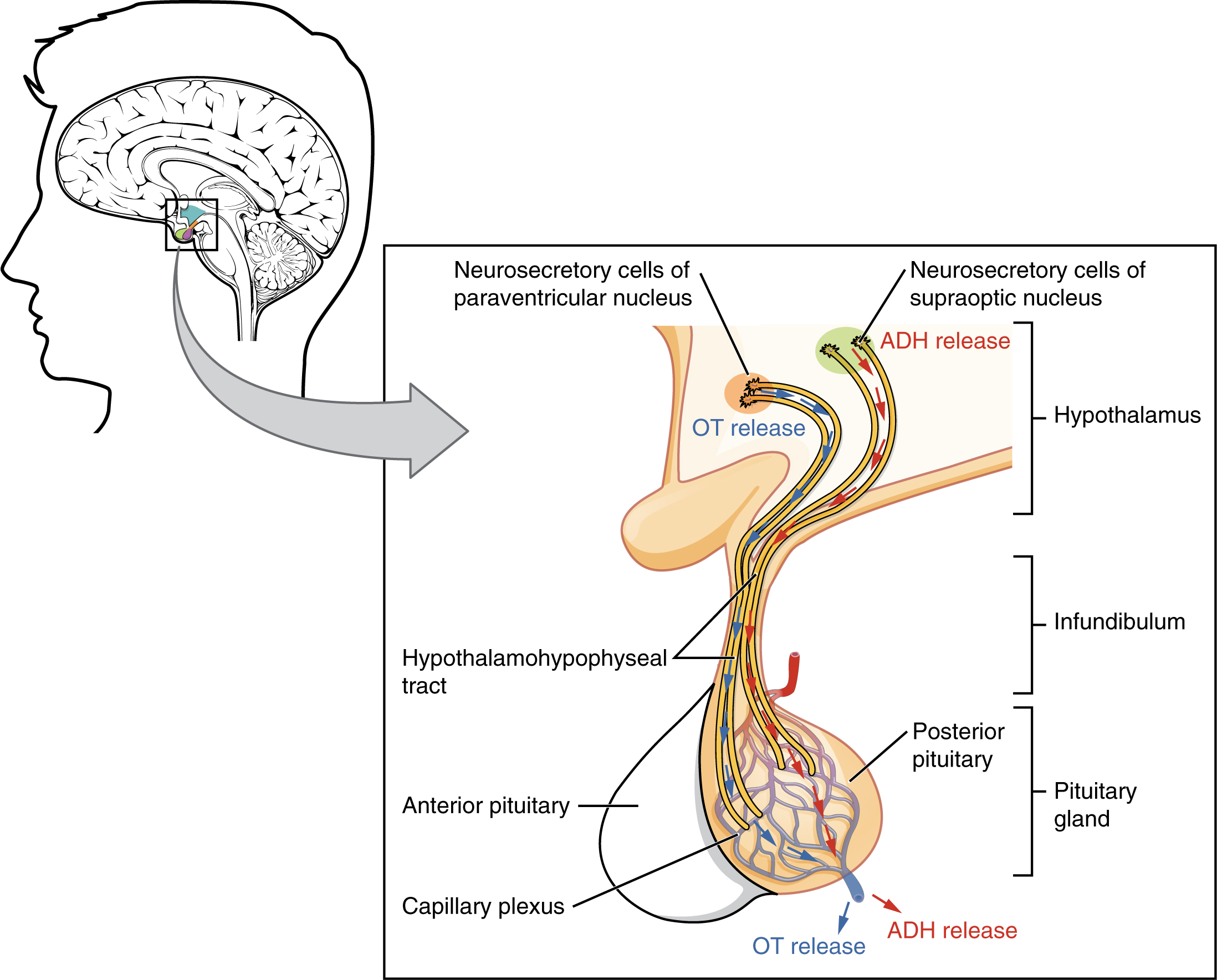
Composed mainly of nerve fibers originating from the hypothalamus, it stores and releases hormones produced in the hypothalamus.
The pituitary gland receives blood from the superior and inferior hypophyseal arteries, branches of the internal carotid arteries. The portal system of veins allows the hypothalamus to directly influence the anterior pituitary.
Function of the Pituitary Gland
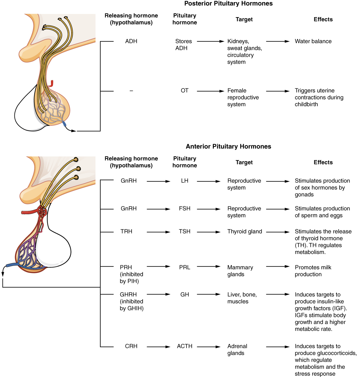
- Anterior Pituitary Hormones: The anterior lobe synthesizes and secretes several hormones, including:
- Growth Hormone (GH): Regulates growth and metabolism.
- Thyroid-Stimulating Hormone (TSH): Stimulates the thyroid gland to produce thyroid hormones.
- Adrenocorticotropic Hormone (ACTH): Stimulates the adrenal glands to produce cortisol.
- Prolactin (PRL): Promotes milk production in the breasts.
- Gonadotropins (LH and FSH): Regulate reproductive processes, including ovulation and spermatogenesis.
- Posterior Pituitary Hormones: The posterior lobe releases:
- Antidiuretic Hormone (ADH): Regulates water balance in the body by controlling kidney function.
- Oxytocin: Stimulates uterine contractions during childbirth and milk ejection during breastfeeding.
Imaging of the Pituitary Gland
- Magnetic Resonance Imaging (MRI):
- Preferred Modality: MRI is the gold standard for imaging the pituitary gland due to its superior soft tissue contrast and ability to delineate small structures within the sella turcica.
- Key Sequences:
- T1-weighted imaging: Provides detailed anatomical information; the normal pituitary appears slightly hyperintense relative to the brain.
- Dynamic contrast-enhanced sequences: Useful for assessing microadenomas and vascular anatomy, as these enhance differently compared to the normal pituitary tissue.
- Visualization: The gland is best visualized in sagittal and coronal planes. MRI can identify gland size, shape, and the presence of any masses or structural anomalies.
- Computed Tomography (CT):
- Role in Imaging: CT is useful for assessing the bony structure of the sella turcica, particularly in cases of trauma or bone abnormalities. However, it is less effective than MRI in visualizing soft tissue detail.
- Indications: CT is typically reserved for situations where MRI is contraindicated or when assessing calcifications or bony erosion associated with pituitary tumors.
4. Clinical Significance
- Pituitary Adenomas: The most common pituitary disorder, adenomas can be classified based on their size:
- Microadenomas: Less than 10 mm, often detected incidentally on MRI.
- Macroadenomas: Greater than 10 mm, may cause symptoms due to mass effect, such as visual disturbances (due to compression of the optic chiasm) or hypopituitarism.
- Hypopituitarism: Can result from pituitary damage due to tumors, surgery, radiation, or trauma. It leads to a deficiency in one or more pituitary hormones, with varied clinical presentations depending on the hormones affected.
- Hyperpituitarism: Excess hormone production, often due to adenomas, can lead to conditions such as acromegaly (excess GH) or Cushing’s disease (excess ACTH).
- Pituitary Apoplexy: A medical emergency caused by hemorrhage or infarction within the gland, leading to sudden headache, visual impairment, and hormonal deficiencies. Imaging is critical for diagnosis and management.
5. Summary
The pituitary gland, though small, is a central player in the endocrine system, controlling a vast array of bodily functions through its hormone production and regulation. Understanding its anatomy and function, alongside its imaging characteristics, is crucial for diagnosing and managing pituitary disorders. MRI remains the cornerstone for pituitary imaging, providing detailed insights into both normal and pathological conditions of this vital gland.
Suggested Further Reading
- Mullur, R., & Liu, J. (2016). The Pituitary Gland: A Clinical Overview. Endocrine Reviews, 37(5), 556-582.
- Bonneville, J.-F., Cattin, F., & Douek, P. (2010). MRI of the Pituitary Gland. Springer-Verlag, Berlin Heidelberg.