Thoracic Cage
The thoracic cage, also referred to as the ribcage, is a complex osseocartilaginous structure that provides protection to vital thoracic and upper abdominal organs while also contributing to respiration. It consists of the sternum, ribs, costal cartilages, and thoracic vertebrae, all of which contribute to the stability, flexibility, and mechanical function of the thorax.
Bony Framework
The thoracic cage is composed of three main skeletal elements:
1. Sternum
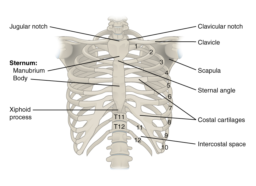
The sternum is a flat, elongated bone located in the anterior midline of the thorax. It consists of three parts:
- Manubrium: The superior portion that articulates with the clavicles at the sternoclavicular joints and the first pair of ribs via costal cartilages.
- Body: The longest part, articulating with the costal cartilages of ribs 2-7.
- Xiphoid Process: The smallest, most inferior portion, which serves as an attachment site for muscles and the linea alba.
The sternum provides anterior support and serves as an important landmark for clinical procedures such as median sternotomy and pericardiocentesis.
2. Ribs
There are 12 pairs of ribs, classified based on their articulation with the sternum:
- True ribs (1-7): Attach directly to the sternum via their costal cartilages.
- False ribs (8-10): Attach indirectly to the sternum via the costal cartilage of the rib above.
- Floating ribs (11-12): Have no anterior attachment to the sternum.
Each rib consists of the following anatomical features:
- Head: Articulates with the corresponding thoracic vertebra and the vertebra above.
- Neck: Lies between the head and the tubercle.
- Tubercle: Articulates with the transverse process of the corresponding thoracic vertebra.
- Shaft: The main body of the rib, curving around the thorax.
- Costal groove: Located on the inferior border of the rib, housing the intercostal neurovascular bundle.
3. Thoracic Vertebrae
The thoracic cage is supported posteriorly by the thoracic spine, which consists of 12 vertebrae (T1-T12). Each thoracic vertebra has characteristic features, including:
- Costal facets on the vertebral bodies for rib articulation.
- Transverse processes that articulate with the rib tubercles.
- Spinous processes that project downward, limiting excessive extension.
Articulations and Ligaments
The thoracic cage has numerous joints that provide stability while permitting respiratory movements.
1. Sternocostal Joints
- Ribs 1-7 articulate with the sternum via their costal cartilages.
- Rib 1 has a synchondrosis with the manubrium, while ribs 2-7 form synovial joints.
2. Costovertebral Joints
- Formed between the rib head and the demifacets of two adjacent thoracic vertebrae.
- Stabilized by the radiate ligament and intra-articular ligament.
3. Costotransverse Joints
- Present in ribs 1-10, where the rib tubercle articulates with the transverse process of the corresponding vertebra.
- Reinforced by the costotransverse ligament and superior costotransverse ligament.
4. Interchondral Joints
- Formed between the costal cartilages of ribs 6-10.
- Provide flexibility to the ribcage.
Musculature of the Thoracic Cage
The thoracic cage serves as an attachment point for numerous muscles involved in respiration, posture, and upper limb movement.
1. Intercostal Muscles
- External intercostals: Elevate ribs during inspiration.
- Internal intercostals: Depress ribs during expiration.
- Innermost intercostals: Assist in rib stabilization.
2. Diaphragm
- The primary muscle of respiration, separating the thoracic and abdominal cavities.
- Contracts to increase thoracic volume, facilitating inspiration.
3. Accessory Muscles
- Scalene muscles: Elevate the first and second ribs.
- Sternocleidomastoid: Elevates the sternum.
- Pectoralis minor and major: Assist in forced inspiration.
Neurovascular Supply
The thoracic cage is supplied by the intercostal neurovascular structures, located within the costal grooves.
1. Intercostal Nerves
- Derived from the ventral rami of thoracic spinal nerves (T1-T11).
- Provide motor innervation to intercostal muscles and sensory innervation to the skin of the thorax.
2. Intercostal Arteries
- Posterior intercostal arteries arise from the thoracic aorta.
- Anterior intercostal arteries arise from the internal thoracic artery.
- Both sets of arteries anastomose within the intercostal spaces.
3. Intercostal Veins
- Drain into the azygos venous system on the right and the hemiazygos system on the left.
Function of the Thoracic Cage
- Protection: Shields the heart, lungs, and major vessels.
- Respiration: Allows expansion and contraction during breathing.
- Support: Provides structural integrity for the thoracic cavity.
Anatomical Variants
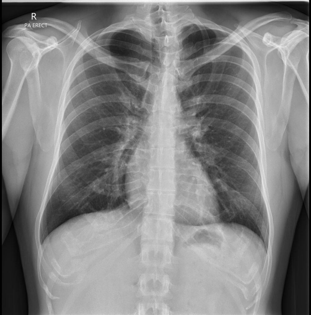
Cervical Rib
- Additional rib arising from C7, seen on the PA view as a bony projection above the first rib.
Bifid or Forked Rib
- A rib with two ossification centres, appearing as a split or forked structure on imaging.
- Typically asymptomatic and incidental.

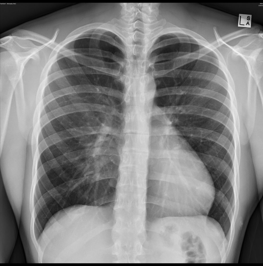
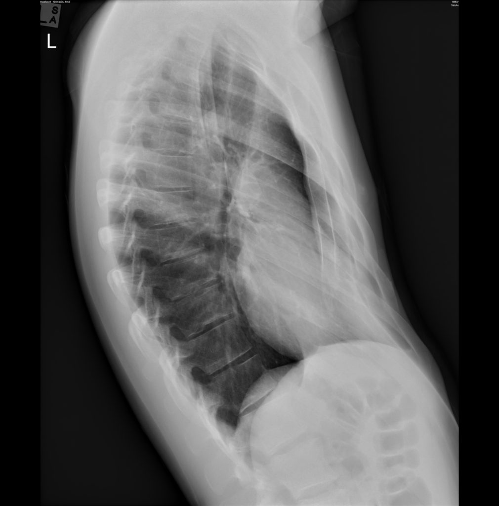
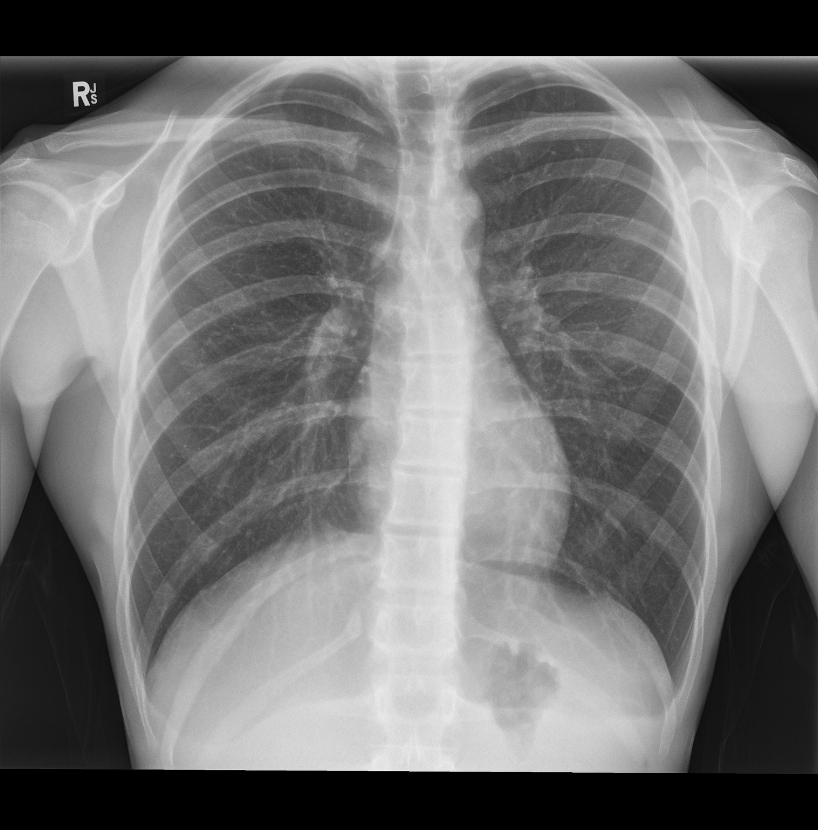
Pectus Excavatum
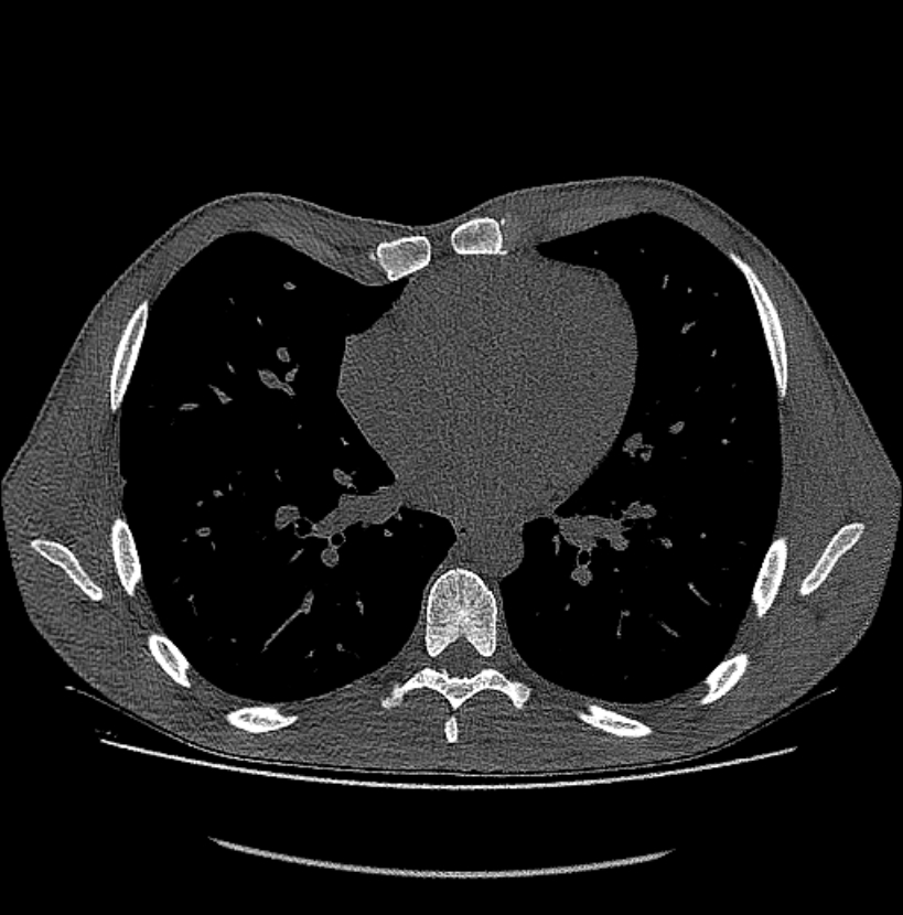
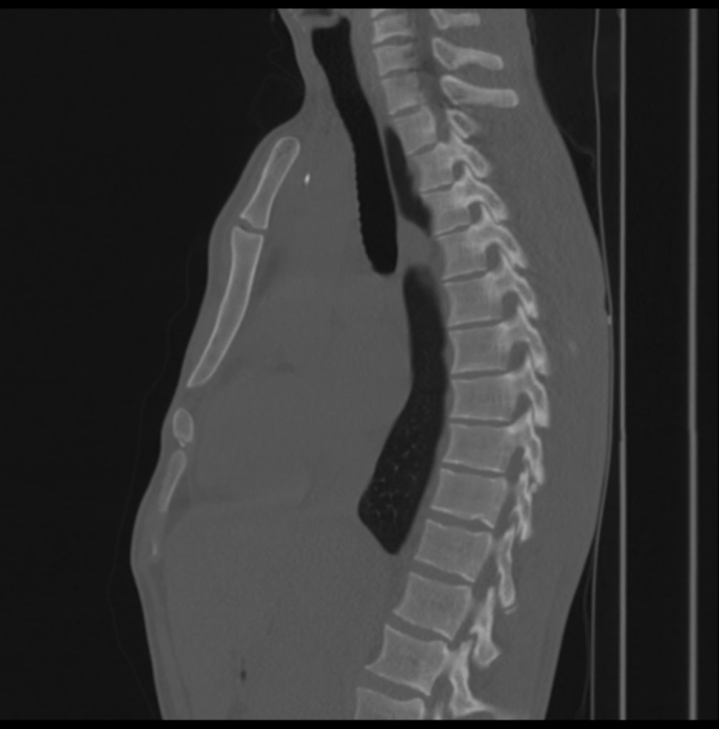
- Concave deformity of the anterior chest wall.
Image findings
- Lateral chest radiograph: Posterior displacement of the sternum.
- PA view: Heart may appear displaced, and ribs slope sharply anteriorly.
- CT: Useful for quantifying severity.
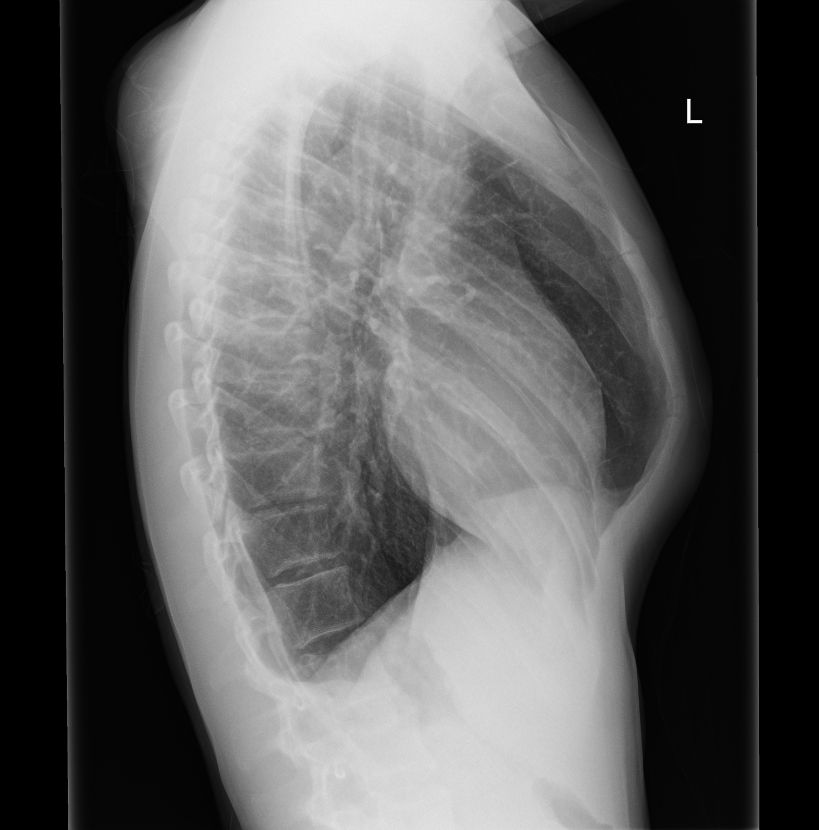
- Pectus Carinatum:
- Protrusion of the anterior chest wall (sternum).
- Radiographs show outward bowing of the sternum with adjacent rib changes.
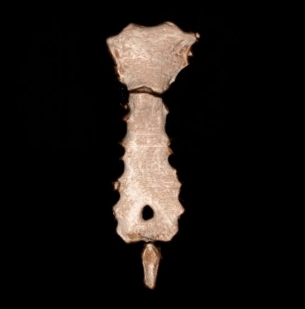
- Sternal Foramen:
- Midline defect in the sternum visible on CT, mimicking lytic lesions.
- Often incidental and of no clinical significance.