

RANZCR Syllabus
Category 1
• Ophthalmic artery
• Infraorbital artery
• Central artery of retina
Category 2
• Supraorbital
• Supratrochlear
• Lacrimal
• Dorsal nasal
• Anterior & posterior ethmoidal
Category 3
• Anterior ciliary
• Posterior ciliary
• Zygomaticotemporal
• Zygomaticofacial
• Ophthalmic artery
• Infraorbital artery
• Central artery of retina
Category 2
• Supraorbital
• Supratrochlear
• Lacrimal
• Dorsal nasal
• Anterior & posterior ethmoidal
Category 3
• Anterior ciliary
• Posterior ciliary
• Zygomaticotemporal
• Zygomaticofacial
Ophthalmic Artery |
|---|
| CATEGORY 1PENDING |
The ophthalmic artery is most commonly the first major intracranial branch of the internal carotid artery. It arises just as the internal carotid artery emerges from the cavernous sinus. The ophthalmic artery has a rich anastomotic network the acts protectively against occlusion.
Origin
C6 segment of the internal carotid artery
Course
The ophthalmic artery has an intracranial and extracranial course.
- It first emerges directly from the C6 segment of the internal carotid artery at the level of the anterior clinoid process of the sphenoid bone.
- It then courses intracranially before passing below the posterior edge of the falciform ligament
- It then pierces the dura mater of the optic nerve and enters the orbit via the optic foramen
- Within the optic canal, the ophthalmic artery travels inferolateral to the optic nerve
- The artery then crosses over (83%) or under (17%) the optic nerve from lateral to medial giving off its first branch
- After emerging from the optic canal, the artery courses anteriorly along the medial wall of the orbit, passing between the superior oblique and lateral rectus muscles
- Along its course, it gives off several branches before dividing into its terminal branches near the upper medial edge of the orbit.
Branches
- Central retinal artery
- Most critical branch of the ophthalmic artery as is fundamental for vision. Most commonly the first branch (78%)
- Supplies the optic nerve as well as the inner layers of the retina
- Muscular branches
- Supplies the extraocular muscles, often via superior and inferior groups
- Accompanies the oculomotor nerve
- Gives off the anterior ciliary arteries
- Posterior ciliary arteries
- Long and short posterior ciliary arteries, pierce the sclera on the posterior aspect of the eyeball lateral to the optic nerve.
- Supplies the choroid and outer layers of the retina
- Lacrimal artery
- A large branch, arising just before the ophthalmic artery enters the orbit
- Supplies the lacrimal gland, eyelids, conjunctiva and parts of the dura mater
- Supraorbital artery
- Arises after the ophthalmic artery crosses over the optic nerve.
- Exits the orbit with the supraorbital nerve and vein through the supraorbital foramen
- Intraorbitally supplies superior rectus and levator palpebrae superioris
- Extraorbitally supplies the skin, muscles and periosteum of the forehead
- Ethmoidal artery (anterior and posterior)
- Anterior is usually larger, it enters the cranial cavity through the anterior ethmoidal foramen and becomes anterior meningeal artery, supplying ethmoidal air cells and the periosteum
- Posterior ethmoid artery supplies the superior oblique, superior and medial rectus and the levator palpebrae superioris
- Medial palpebral arteries
- Terminal branches: supratrochlear artery, dorsal nasal artery
Supply
All structures of the orbit, upper two-thirds of the nose, nasal cavity, frontal area of the scalp, ethmoid and frontal sinuses, parts of meninges
Variants
- Ophthalmic artery originating from the middle meningeal artery
- Ophthalmic artery originating from anterior cerebral artery
- Lacrimal artery originating from the middle meningeal artery
- Persistent dorsal ophthalmic artery
- Persistent ventral ophthalmic artery
Infraorbital Artery |
|---|
| CATEGORY 1PENDING |
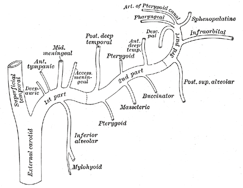
The infraorbital artery is a branch of the third part of the maxillary artery.
Short notes on the Maxillary Artery
The maxillary artery branches off the external carotid artery and passes through the infratemporal fossa. It then courses upward and medial to the mandibular joint toward the maxillary bone. It then runs along the pterygopalatine fossa and enters the orbit through the inferior orbital fissure as the infraorbital artery.
Origin
Maxillary artery – Third part
- Continues from the trunk of the maxillary artery in conjunction with the posterior superior alveolar artery
Course
- Arises from the maxillary artery
- Courses along the infraorbital groove of the maxillary bone before passing through the infraorbital canal with the infraorbital nerve
- It exits through the infraorbital foramen beneath the infraorbital head of the levator labii superioris muscle
Branches
- orbital branches – assist in supplying the inferior rectus muscle, inferior oblique muscle and the lacrimal sac
- anterior superior alveolar arteries – descends through the anterior alveolar canals to supply the upper incisor and canine teeth and the mucous membrane of the maxillary sinus.
Supply
- Lower eyelid
- Lacrimal sac
- Anastomoses with the angular and dorsonasal artery
References
Michalinos, A., Zogana, S., Kotsiomitis, E., Mazarakis, A. and Troupis, T., 2015. Anatomy of the ophthalmic artery: a review concerning its modern surgical and clinical applications. Anatomy research international, 2015.