

There are two major vessels of the coronary circulation that branch to supply the entire heart, named left and right coronary arteries. Both arteries arise from their respective coronary sinus (or ostia), which are small openings found behind the left and right flaps of the aortic valve.
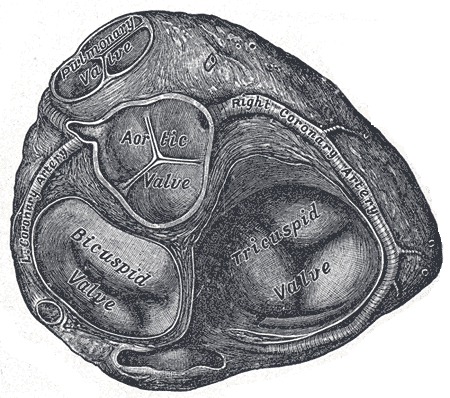
Left Coronary Artery
Origin
Left coronary sinus (of Valsava), superior to the aortic root.

Course & Relations
- The left main coronary artery (LCA) passes posteriorly to the pulmonary trunk
- Descends into the left coronary sulcus between pulmonary trunk and auricle of the left atrium
- Bifurcates into terminal branches just before entering coronary sulcus (atrioventricular groove)
Branches
- Left anterior descending branch (LAD)
- Continues in the same direction as the LCA, i.e. 180 degrees
- Descends down the anterior surface of the heart in the anterior interventricular sulcus along with the great cardiac vein, towards the apex of the heart
- Gives a number of diagonal arteries, supplying the left ventricle
- Gives perforating septal branches, which branch at 90 degrees to the LAD to supply the anterior 2/3 of the interventricular septum.
- Left circumflex artery (LCx)
- Follows coronary sulcus toward the posterior surface of the heart
- Gives obtuse marginal branch to supply obtuse marginal surface
- Usually diminishes before reaching posterior interventricular sulcus
- Left marginal artery (LMA)
Variants
- Left dominant variant, large left circumflex supplies posterior interventricular artery
- Left coronary artery supplying SA nodal branch (40%)
- LCx supplying AV node
- Coronary artery arising from non-coronary sinus
- Co-dominance of the arteries
- Ramus intermedius – an additional artery arising at the bifurcation of the left main artery
Right Coronary Artery
Origin
Right coronary sinus (of Valsalva), superior to the aortic root.
Course & Relations
- Descends into coronary sulcus between auricle of the right atrium and pulmonary trunk
- Associated with small cardiac vein
- Gives branch that passes posteriorly to the right of the aortic root, curves around posterior to superior vena cava to supply sino-atrial node
- Continues in coronary sulcus toward posterior surface of the heart
- Gives acute marginal branch to inferior margin of the heart
- Near posterior interventricular sulcus, gives AV nodal branch
- Turns to continue down posterior interventricular sulcus as the posterior interventricular artery
- Supplies the inferior wall, posterior 1/3 of the interventricular septum, and the posteromedial papillary muscle.
- Associated with middle cardiac vein
Branches
- Conus branch: suppliues RV outflow tract
- Sinoatrial nodal branch
- Right marginal branch (supplies RV free wall and inferior margin of heart and apex)
- Posterior interventricular branch
- AV nodal branch
Variants
- Right dominance: RCA gives of posterior descending artery (80%)
- Left dominance/Co-dominance: LCA gives PDA or both contribute (20%)
- RCA supplies SA nodal artery (60%)
- LCx supplies SA nodal (40%)
- Anomalous courses of the RCA e.g. origin from the left aortic sinus (rare)