

Overview
The aorta is the largest artery in the body. It receives cardiac output from the left ventricle and supplies the body with oxygenated blood via the systemic circulation.
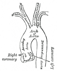
Origin
Originates from the left ventricle of the heart, anterior to the pulmonary artery.
Segments & Course
Aortic root
- First part of the aorta, located between the aortic annulus (junction of left ventricular outflow tract and aortic valve) and the sinotubular junction (divides the aortic root from the ascending aorta)
- It has several subparts:
- three aortic valve leaflets and leaflet attachments
- three aortic sinuses of Valsalva – intraluminal space formed by the cusps
- right and left contain coronary artery ostia, with a third non-coronary aortic sinus located posteriorly
- three interleaflet trigones: space between the cusps and the annulus
- Average aortic diameter: 3.0 cm ± 0.5
Ascending aorta
- From the sinotubular junction, the aorta ascends into the superior mediastinum i.e. above the transthoracic (of Ludwig) plane
Aortic arch
- The aorta then arches posteriorly over the left lung root, to the left of the vertebral bodies
- The arch ends as it enters the posterior mediastinum
- Conventionally, the aortic arch gives off three branches (in order):
- Brachiocephalic trunk (innominate artery)
- Left common carotid artery
- Left subclavian artery
- Distal to the origin of the left subclavian artery is a section known as the aortic isthmus where there is a mild narrowing of the aorta which occurs at the site of the ligamentum arteriosum (remnant of the ductus arteriosus)
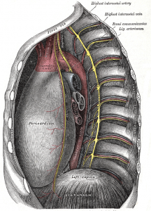
Descending aorta
- From the transthoracic plane, the aorta descends in the posterior mediastinum to the diaphragmatic hiatus at the level of T12 where it exits the thorax and becomes the abdominal aorta
- The descending aorta gives off multiple vessels before exiting the thoracic cavity:
- arteries to supply the pericardium, bronchi, mediastinum, and esophagus
- superior phrenic arteries
- posterior intercostal arteries
- subcostal arteries
Development
- Develops during third gestational week
- The ascending aorta and aortic arch develop independently from two different embryologic tracts
- Ascending aorta develops from the truncus arteriosus
- Aortic arch forms through the development of the branchial arch arteries
Variants
- Double aortic arch – forms vascular ring around trachea and oesophagus, then joins as single descending aorta
- Right sided arch – passes to right or trachea, usually associated with mirror image branching of left subclavian
- Bovine arch – common origin of brachiocephalic artery and left common carotid artery
- Left vertebral artery originates from the arch between left common carotid and subclavian
- Aberrant right subclavian arising distal to left subclavian
- Left brachiocephalic artery
- Right common carotid and subclavian arise independently
- Thyroid ima artery arising between brachiocephalic and left common carotid artery
References
Anatomy, Thorax, Heart Aorta
https://www.ncbi.nlm.nih.gov/books/NBK538140/
https://www.ncbi.nlm.nih.gov/books/NBK538140/