

Overview
Large, wide, flat C-shaped white matter tract connecting the left and right cerebral hemispheres. It forms the floor of the longitudinal fissure and part of the roof of the lateral ventricles.

[foogallery id=”7053″]
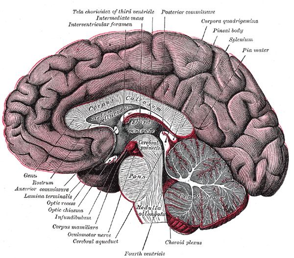
Structure
Rostrum
- Beak-shaped (Rostrum – “Bird’s beak”)
- Connect the orbital surfaces of the frontal lobes
- Continuous with the lamina terminalis – a thin sheet of gray matter and pia mater that attaches to the upper surface of the chiasm and stretches upward to fill the interval between the optic chiasm and the rostrum of the corpus callosum
Genu
- Bend of the anterior corpus callosum (Genu – “knee”)
Body (or trunk)
- Long central section, curves posteroinferiorly infront of the septum pellucidum.
- Connects the temporal lobes of the hemispheres as well as the occipital lobes
- Connected to the fornix by the septum pellucidum
Splenium
- Posterior part
- Connects the posterior cortices, predominantly parietal and temporal areas
Connections
- Forceps minor (anterior forceps) – connects the medial and lateral surfaces of the frontal lobes via the genu
- Forceps major (posterior forceps) – connects occipital lobes via splenium
- Thicker axons in the trunk of the corpus callosum interconnect areas of the motor cortex
- The splenium communicates somatosensory information between the two halves of the parietal lobe and the visual cortex
Arterial Supply
Majority of arterial supply is derived from branches of the anterior cerebral artery and posterior cerebral artery:
- Pericallosal artery – distal part of anterior cerebral artery, courses along the superior surface of body of corpus callosum in pericallosal cistern
- Posterior pericallosal artery (also known as splenial artery) – usually a branch of the posterior cerebral artery
80% of patients may have additional supply from the anterior communicating artery via:
- Subcallosal artery – supplies hypothalamus and the medial portions of rostrum and genu
- Median callosal – supplies hypothalamus, the medial portion of rostrum and genu as well as the body
Venous Drainage
Callosal veins and callosocingulate veins towards the deep venous system of the brai
Development
- Begins to form around week 12
- Begins with genu and continues posteriorly along the body to the splenium. The rostrum forms last.
- Myelination occurs in the opposite direction, from the splenium forward
Relations
- Superiorly: interhemispheric fissure, falx cerebri, anterior cerebral vessels
- Inferiorly: septum pellucidum, fornix, lateral ventricles, choroid plexus within lateral ventricles, thalamus
- Anteriorly: dentate gyrus
- Posteriorly: Fornix and commissure
- Deep to the cingulate gyrus and separated by the callosal sulcus
- The anterior cerebral arteries are in contact with the under surface of the rostrum
Anatomical Variants
- Agenesis, hypogenesis (partial formation), hypoplasia (under development, e.g. too thin), dysgnesis (malformation)
- Callosal isthmus
- the site of interconnecting axons from the left and right superior temporal and parietal lobes, located at the junction of the posterior body and splenium
- appears relatively thin relative to the other parts of the corpus callosum
- can mimic focal atrophy of the corpus callosum as seen in cerebral palsy, demyelinating disorders, trauma or leukodystrophies.
- Callosal vascular indentation
- usually from an A3 segment of the anterior cerebral artery
- common at the posterior genu or anterior body
- can mimic a congenital anomally – callosal hypensis or atrophy
- Callosal-septal and pericallosal pseudolesions
- midline sagitally FLAIR images may demonstrate a normal linear hyperintense signal at the callosal-septal interface
- can mimic multiple sclerosis, however, this signal is oriented parallel to the lateral ventricle, whereas early demyelinating lesions of multiple sclerosis are oriented perpendicular to the curve of the corpus callosum and lateral ventricles
- Subcallosal/paraterminal pseudomass
- midline sagittal images (particularly FLAIR images) occasionally demonstrate a pseudomass immediately below the rostrum and anterior to the third ventricle
- due to volume averaging of the paraterminal/subcallosal gyri near the midline (i..e. not present on axial or coronal images)
- can mimic a meningioma, carinopharyngioma, optic glioma, hypothalamic glioma or aneurysm
References
McKinney A.M. (2017) Midline Variants of the Septum Pellucidum, Corpus Callosum, and Massa Intermedia. In: Atlas of Normal Imaging Variations of the Brain, Skull, and Craniocervical Vasculature. Springer, Cham.