

Overview
The ventricules of the brain produces and transmits cerebrospinal fluid into the ventricular system, subarachnoid space and spinal canal.
The fourth ventricle is the most inferiorly located ventricle

Boundaries
- Roof (tent-shaped)
- Superior part – Superior medullary velum, a think lamina connecting the left and right superior cerebellar peduncles together
- Inferiorly part – Inferior medullary velum
- Superolaterally – bordered by the superior cerebellar peduncle
- Inferolaterally – bordered by the inferior cerebellar peduncles and the teniae – meets inferiorly at the midline to form the obex
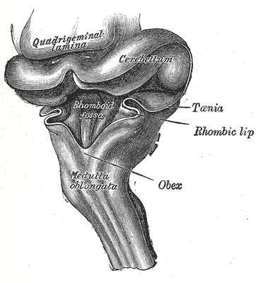
- Floor (anterior surface) – Rhomboid fossa. The fossa consists of three parts, superior, intermediate, and inferior:
- Superior part – Triangular in shape and limited laterally by the superior cerebellar peduncle; its upwardly directed apex is continuous with the cerebral aqueduct; its base is represented by an imaginary line at the level of the upper ends of the superior foveae.
- Intermediate part – Extends from this level to that of the horizontal portions of the taeniae of the ventricle; it is narrow above where it is limited laterally by the middle peduncle, but widens below and is prolonged into the lateral recesses of the ventricle.
- Inferior part – Triangular with a downwardly directed apex, named the calamus scriptorius (as is shaped like a writing quill-nib), which is continuous with the central canal of the closed part of the medulla oblongata.

Features
- Median sulcus – divides the rhomboid fossa into left and right halves, extends the length of the ventricle
- Sulcus limitans – further divides each half, forms the lateral boundary of the medial eminence.
- Medial eminence – elevation between the median sulcus and sulcus limitans
- Stria medullaris – fibers originating from arcuate nuclei, runs transversely across the floor into inferior cerebellar peduncles at the widest part of rhomboid fossa
- Above stria medullaris:
- Medial eminence – longitudinal elevation on either side of median sulcus
- Sulcus limitans – at the lateral border of medial eminence
- Superior fovea – sulcus limitans widens superiorly into a triangular depression, locus ceruleus lies just superior
- Vestibular area – area between sulcus limitans and the lateral angles, overlies vestibular nuclei
- Facial colliculus – bilateral swelling on the medial eminence at the level of superior fovea, formed by fibers from motor nucleus of facial nerve as they hook around abducen nucleus
- Below stria medullaris:
- Medial eminence divided into upper hypoglossal trigone and a lower vagal trigone by a sulcus
- Funiculus separans – a narrow ridge across vagal triangle
- Area postrema – a small area below funiculus separans and vagal triangle
Contents
- Tela choroidea – made up of two layers of pia mater
- Choroid plexus – a highly vascular structure which lies within the folds of pia mater forming the tela choroidea.
These structures are responsible for the production of the cerebrospinal fluid, which has several roles:
- Fills the subarachnoid space and ventricles
- Plays a protective role to the brain
- Supplies nutrients to the brain
- Removes waste products from the brain, which are absorbed by the arachnoid villi.
Arterial Supply
Anterior inferior cerebellar artery (AICA), the posterior inferior cerebellar artery (PICA) and the superior cerebellar artery (SCA) are the main supplying vessels.
- AICA supplies the portion of the plexus in the cerebellopontine angles and the adjacent part of the lateral recess of the fourth ventricle through the foramina of Luschka.
- PICA supplies most of the choroid plexus in the roof and the median opening of the fourth ventricle.
CSF Tributaries
- Superiorly – The fourth ventricle connects to the third ventricle through a thin canal called the cerebral aqueduct (of Sylvius).
CSF Drainage
There are four routes via which cerebrospinal fluid may flow from the fourth ventricle:
- Inferiorly – CSF drains via obex into the central canal of the spinal cord
- Superolaterally – CSF drains into two lateral apertures called the foramina of Luschka which helps spread CSF to the subarachnoid cisterns, which bathes the brain, between arachnoid mater and pia mater. Here the CSF is reabsorbed back into the circulation.
- Inferomedially – CSF drains into the median aperture called the foramen of Magendie which drains to the subarachnoid cisterns
Relations
Anteriorly
- Pons
- Medulla
Posteriorly
- Cerebellum
Inferiorly
- Spinal cord and canal
- Cuneate (lateral) and gracile (medial) tubercules