

• Lateral ventricles
• Third ventricle & boundaries
• Cerebral aqueduct
• Fourth ventricle
• Obex, median (Magendie) and lateral (Luschka) foramina
• Choroid plexus
Category 2
• Septum pellucidum, velum interpositum
• Choroid fissures of lateral ventricles
• Superior medullary velum
• Features of fourth ventricle floor
Category 3
Nil
The ventricular system is a set of four interconnected cavities, each containing choroid plexuses which produce cerebrospinal fluid. The ventricular system is continuous with the central canal of the spinal cord from the fourth ventricle.

Lateral Ventricles |
|---|
| CATEGORY 1PENDING |
The pair of lateral ventricles are the largest of the ventricles, located in each cerebral hemisphere. Each lateral ventricle is a C-shaped cavity when viewed laterally,
Boundaries
- Commences at the inferior horn in the temporal lobe and terminating at the interventricular foramina (foramen of Monro) where each ventricle connects to a single midline third ventricle.
- The posterior horn extends posteriorly into the occipital lobe.
- The anterior horn extends into the frontal lobe.

Structure
- Anterior (frontal) horn: Extends into the frontal lobe and connects to the third ventricle via the interventricular foramen.
- The roof is formed by the anterior part of the trunk of the corpus callosum
- The floor is formed by the head of the caudate nucleus.
- The medial wall is formed by the septum pellicudum, which separates the two anterior horns.
- Body: The central part of the ventricle, elongated anteroposteriorly between the anterior horn and the trigone.
- The roof is bound by the trunk of the corpus callosum. The tail of the caudate nucleus form the upper portion of the lateral edge.
- The medial wall is formed by the septum pellucidum and body of the fornix. The body reaches the splenium of the corpus callosum.
- The floor is formed by the superior surface of the thalamus medially and caudate nucleus laterally.
- Trigone: The confluence of the occipital and temporal horns.
- Inferior (temporal) horn: It begins at the posterior end of the central region and runs anteroinferiorly into the temporal lobe.
- The roof is formed by the tapetum of the corpus callosum and cauda of the caudate nucleus.
- The lateral wall is formed by fibres of the tapetum
- The medial wall is formed by the caudate nucleus and the stria terminalis.
- The floor is formed by the hippocampus, alveus and fimbriae.
- Posterior (occipital) horn: Extends posteromedially into the occipital lobe
- The roof is formed by the tapetum of the corpus callosum
- The lateral wall is formed by tapetum
- The medial wall is formed in part by the forceps and white matter of the occipital lobe cortex
Third Ventricle |
|---|
| CATEGORY 1PENDING |
A midline funnel-shaped cavity, comprising the central part of the ventricular system.
Boundaries
- Roof: Extends from the foramen on Monro anteriorly to the suprapineal recess posteriorly. Lies immediately inferior to the body of fornix.
- Floor: Extends from the optic chiasm anteriorly to the aqueduct of Sylvius posteriorly
- Formed by the optic chiasm, infundibulum of the hypothalamus, tuber cinereum, mammillary bodies, posterior perfortate substance, anterior part of the tegmentum of the midbrain.
- Anterior wall: Extends from the foramen of Monro superiorly to the optic chiasm inferiorly
- Formed by the foramen of Monro, columns of fornix, anterior commissure, lamina terminalis, optic recess, optic chiasm
- Posterior wall: Extends from the suprapineal recess superiorly to the aqueduct of Sylvius inferiorly.
- Formed by the suprapineal recess, habenular commissure, pineal body and its recess, posterior commissure, aqueduct of Sylvius
- Lateral walls: Formed by the medial aspects of the thalamus and hypothalamus which are separated by the hypothalamic sulcus
Communications
- Foramen of Monro: communication with each lateral ventricle
- Aqueduct of Sylvius: communication with the fourth ventricle
Recesses
- Supra-optic recess: located superior to the optic chiasm and inferior to the lamina terminalis
- Infundibular recess: lies on the floor, between the optic chiasm and mamillary bodies. It extends inferiorly into the pituitary stalk
- Pineal recess: projects posteriorly into the pineal body, between the cranial and caudal lamina of the pineal gland
- Suprapineal recess: located between the superior portion of the pineal gland and the inferior aspect of the tela choroidea in the roof. It extends posteriorly, behind the posterior wall.
Fourth Ventricle |
|---|
| CATEGORY 1PENDING |
The fourth ventricle has a characteristic diamond shape named the rhomboid fossa and posterior tent-shaped roof. It is located within the pons or upper part of the medulla oblongata.

Boundaries
- Extends from the cerebral aqueduct (Aqueduct of Sylvius) superiorly
- Extends into the central canal of brainstem and spina cord
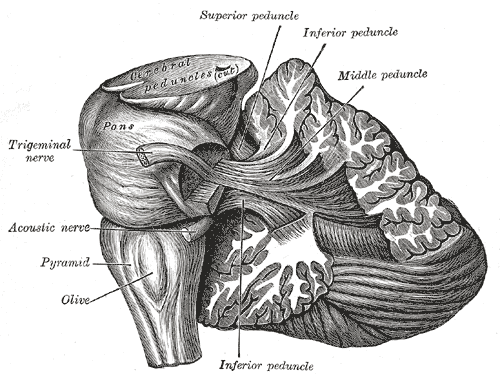
- Lateral walls: formed by the cerebellar penducles
- The superior lateral wall formed by the superior cerebellar peduncle
- The inferior lateral wall formed by the inferior cerebellar peduncle, the gracile and cuneate tubercles
- It features a pair of lateral recesses either side of the midline known as the foramina of Luschka
- Roof (posterior surface)
- tent-like structure, peaking posteriorly as the fastigium and extending into the white core of the cerebellum
- formed by the superior and inferior medullary velum (thin lamina connecting the left and right cerebellar peduncles) and superior cerebellar penduncle
- Floor (anterior surface)
- formed by the rhomboid fossa
- features a median sulcus extending the length of the ventricle that divides the floor into left and right halves
- the sulcus limitans further divides the halves
- Motor neurons are located medial to the sulcus limitans, whereas sensory neurons are located laterally
- the rhomboid fossa can also be divisible by the striae medullares into a superior and inferior triangle
Relation
- Posterior to the pons and medulla
- Anterior to the cerebellum
Communications
- Cerebral aqueduct
- The cavity of the fourth ventricle communicates with the third ventricle superiorly as a continuation of the cerebral aqueduct
- Lateral apertures (foramina) of Luschka
- Lateral recesses which extend between the inferior cerebellar peduncle and the peduncle of the flocculus of the cerebellum
- Opens into the subarachnoid space via the cistern of great cerebral vein
- specifically into the pontocerebellar cistern at the cerebellopontine angle.
- Media aperture of Magendie
- Opens into subarachnoid space via the cisterna magna
- Obex
- The cavity of the fourth ventricle communicates with the central canal of the brainstem (and latter the verterbral column).
Choroid Plexus |
|---|
| CATEGORY 1PENDING |
The choroid plexus (or plica choroidea) consists of modified ependymal cells surrounding a core of capillaries which arise from the tela choroidea in each of the cerebral ventricles. The choroid plexus produces and secrete the cerebrospinal fluid.
The tela choroidea is a thin, highly vascularized, loose connecting region of the pia mater that adheres to the underlying ependyma.
Location
The choroid plexus resides in the pia mater (innermost meningeal layer).
- Lateral ventricle: the floor of the body, the roof of the temporal horns
- Anterior horn: there is no choroid plexus
- Third ventricle: roof, extending into the interventricular foramen
- Fourth ventricle: a small T-shaped structure, indenting the medullary part of the roof
Blood
- Anterior choroidal branch of the internal carotid artery and choroidal branches of the posterior cerebral artery
Venous drainage
- Single choroidal vein