

• Cornea & sclera
• Choroid & retina
• Iris & lens
Category 2
Nil
Category 3
• Canal of Schlemm
• Macula position
• Short ciliary arteries
• Nerve supply
– Short and long ciliary nerves
• Ciliary ganglion
Cornea & Sclera |
|---|
| CATEGORY 1PENDING |
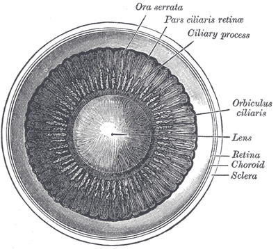 The cornea and sclera constitute the outer layer of the eye.
Cornea
Transparent avascular tissue covering the iris, pupil and anterior chamber. Its primary function is to refract light, accounting for two-thirds of the eye’s total optical power.
Layers
- Corneal epithelium – thin layer of epithelial
- Bowman’s layer – tough layer of collagen
- Corneal stroma – thick, transparent middle layer
- Descemet’s membrane – thin acellular layer serving as a modified basement membrane of the corneal endothelium
- Corneal endothelium – simple think layer of mitochondria rich cells
Innervation
- Densely innervated with sensory nerve fibres via the ophthalmic branch of the trigeminal nerve
Sclera
The opaque white, fibrous outer layer of the eye. It forms more than 80% of the surface area of the eyeball extending from the cornea all the way to the optic nerve and is continuous with the dura mater posteriorly. Anteriorly it is continuous with the limbus of the cornea. It provides protection to the eye and an attachment for the insertion of extraocular muscles.
Choroid & Retina |
|---|
| CATEGORY 1PENDING |
Choroid
The choroid is a highly vascular layer of the eye and lies between the retina and sclera. It is important to the nutrient and oxygen exchange with the outer retina.
Blood supply
- Uveal circulation
- Derived from branches of the ophthalmic artery
- Supplies the uvea, middle and outermost layers of the retina
- Retinal circulation
- Derived from the central retinal artery
- Supplies
Posterior ciliary arteries (from the ophthalmic artery)
mater posteriorly. Anteriorly it is continuous with the limbus of the cornea. It provides protection to the eye and an attachment for the insertion of extraocular muscles.
Iris & Lens |
|---|
| CATEGORY 1PENDING |
Iris
Pigmented muscular curtain that lies between the cornea and lens, forming a circular aperture termed the pupil.
Layers
- Stroma – front pigmented fibrovascular layer, connected to the sphincter pupillae which contracts the pupil and dilator pupillae which enlarges the pupils
- Pigmented epithelial cells