

Overview
A complex wedge-shaped space posterolateral to the maxillary sinus, below the skull base and between side wall of pharnynx and mandible. It acts as conduit for neurovascular structures entering and leaving cranial cavity. It is associated with the muscles of mastication, pterygoplatine fossa and temporal fossa.
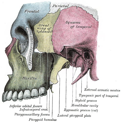
Borders
- Anterior – posterolateral wall of the maxillary sinus
- Posterior – carotid sheath
- Medial – lateral surface of lateral pterygoid plate, tensor veli palatini and levator veli palatini muscles, superior constrictor muscle
- Lateral – coronoid process and ramus of mandible, zygmoatic arch
- Roof – greater wing of sphenoid, foramen ovale, foramen spinosum
- Floor – medial pterygoid muscle
Contents
- Deep part of the parotid gland
- Muscles
- Medial and lateral pterygoid muscle
- Inferior part of the temporalis muscle
- Nerves:
- Mandibular nerve (CN VIII) – Enters fossa via foramen ovale. Gives rise to motor and sensory branches (inferiorly innervates cutaneous structures of the face).
- Auriculotemporal, buccal, lingual and inferior alveolar nerves – sensory branches of the trigeminal nerve.
- Chorda tympani (branch of CN VII) – follows the anatomical course of the lingual nerve and provides taste innervation to the anterior two-thirds of the tongue.
- Otic ganglion
- Vessels:
- Maxillary artery – terminal branch of the external carotid artery. Passes through the pterygomaxillary fissure, enters the pterygopalatine fossa in front of the ganglion and gives off five branches
- Within the fossa, it gives rise to the middle meningeal artery, which passes through the superior border via the foramen spinosum.
- Pterygoid venous plexus– drains the eye and is directly connected to the cavernous sinus.
- It provides a potential route by which infections of the face can spread intracranially.
- Maxillary vein
- Middle meningeal vein
- Maxillary artery – terminal branch of the external carotid artery. Passes through the pterygomaxillary fissure, enters the pterygopalatine fossa in front of the ganglion and gives off five branches
Communications
- Middle cranial fossa – via the foramen ovale and foramen spinosum.
- Temporal fossa – via the space deep to the zygomatic arch
- Pterygopalatine fossa – via the pterygomaxillary fissure