

Overview
The muscles of mastication are associated with movements of the mandible at the temporomandibular joint.

Muscles
Masseter
- Description: powerful muscle covering lateral aspect of mandibular ramus, composed of superficial and deep head
- Origin: zygomatic arch and maxilla
- Insertion: angle and ramus of mandible
- Action: primer mover of jaw closure; elevates mandible
- Relations:
- Lateral to mandible
- Nerve supply: Mandibular division of the trigeminal nerve
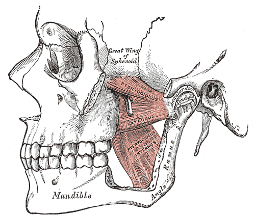
Lateral Pterygoid
- Description: deep two-headed muscle
- Origin: Greater wing and lateral pterygoid plate of sphenoid bone
- Insertion: condyle of mandible and capsule of temporomandibular joint
- Action: protrudes mandible, side-to-side grinding
- Relations:
- Lies superior to medial pterygoid muscle
- Defines segments of the maxillary artery. 1st mandibular part – posterior to lateral pterygoid. 2nd pterygoid part – within lateral pterygoid. 3rd pterygopalatine part – anterior to lateral pterygoid
- Nerve supply: Mandibular division of the trigeminal nerve
Medial pterygoid
- Description: deep two-headed muscle running along internal surface of mandible
- Origin: medial surface of lateral pterygoid plate of sphenoid bone, maxilla and palatine bone
- Insertion: Medial surface of mandible near its angle
- Action: assists in elevation of mandible, protrudes mandible
- Nerve supply: Mandibular division of the trigeminal nerve
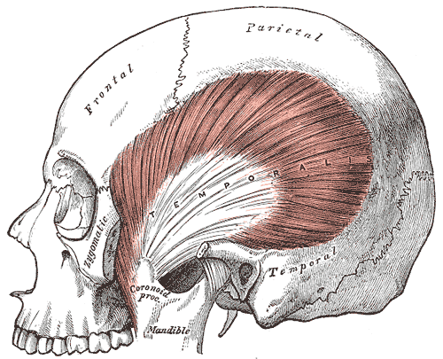
Temporalis
- Description: fan-shaped muscle covering temporal, frontal and parietal bones
- Origin: temporal fossa
- Insertion: coronoid process of mandible
- Action: closes jaw, elevates and retracts mandible
- Nerve supply: Mandibular division of the trigeminal nerve
Development
The muscles of mastication develop from the first pharyngeal arch. Thus, they are innervated by a branch of the trigeminal nerve (CN V), the mandibular nerve.
References
Marieb, E.N. and Hoehn, K., 2007. Human anatomy & physiology. Pearson education. Vancouver