

Overview
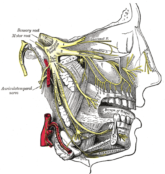
The trigeminal nerve is the fifth and largest cranial nerve (CN V), involved in facial sensation and motor supply to the muscles of mastication.
Origin
Originates from three sensory nuclei (mesencephalic, principal sensory, spinal nuclei of the trigeminal nerve) and one motor nucleus (motor nucleus of the trigeminal nerve).
Sensory
- Mesencephalic nucleus – proprioceptive fibres for muscles of the face, orbit, mastication, and tongue; extends through the whole length of the midbrain, lateral to the cerebral aqueduct
- Principal nucleus – located in the upper pons, lateral to the motor nucleus. Responsible for touch sensation for all three trigeminal divisions.
- Spinal nucleus – lower pons to upper cervical cord, is responsible for pain and temperature; additionally it receives afferent fibres from the glossopharyngeal nerve and vagus nerve.
Motor
- The motor nucleus is located in the upper pons and gives off the smaller motor root which bypasses the trigeminal ganglion and innervates the muscles of mastication as well as mylohyoid, the anterior belly of digastric, tensor tympani and tensor palatini.

Course
- At the level of the pons, the sensory nuclei merge to form a sensory root and the motor nucleus continues to form a motor root.
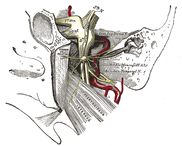
- In the middle cranial fossa, the sensory root expands into the trigeminal ganglion (semilunar or Gasserian ganglion) located lateral to the cavernous sinus, in a depression called the trigeminal (Meckel’s) cave, entering via the porus trigeminus.
- The trigeminal ganglion gives rise to 3 divisions: ophthalmic (V1), maxillary (V2) and mandibular (V3).

- The motor root passes inferiorly to the sensory root, along the floor of the trigeminal cave. Its fibres are only distributed to the mandibular division.
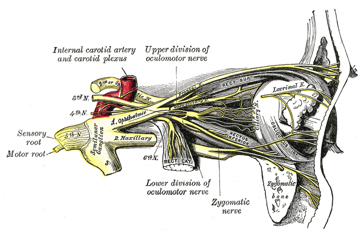
- The ophthalmic nerve and maxillary nerve travel within the lateral wall of the cavernous sinus exiting the cranium via the superior orbital fissure and foramen rotundum respectively.
- The mandibular nerve exits via the foramen ovale entering the infratemporal fossa.
 Divisions
Ophthalmic nerve (CN V1) – Superior division
- Skull exit: Superior orbital fissure
- Sensory supply (only):
- Frontal branch: largest, runs along roof of orbit toward opening, bifurcating into cutaneous supraorbital and supratrochalear nerves (forehead and scalp)
- Nasociliary branch: only branch that travels through the Annulus of Zinn and supplies branches to the eyeball and divides into posterior ethmoidal, anterior ethmoidal and infratrochlear. The anterior and posterior ethmoidal nerves terminate at the external nasal nerve (external nerve). The infratrochlear nerve is a terminal branch of the nasociliary nerve.
- Lacrimal branch: smallest, primarily cutaneous branch but indirectly associated with innervation of the lacrimal gland
Maxillary nerve (CN V2) – Intermediate division
- Skull exit: Foramen rotundum (base of greater wing of sphenoid)
- Course:
- After arising from the trigeminal ganglion in Meckel’s cave (within middle cranial fossa) the maxillary nerve passes through the lateral wall of the cavernous sinus below the ophthalmic nerve
- It then leaves the middle cranial fossa through the foramen rotundum and enters the pterygopalatine fossa
- The main trunk leaves the pterygopalatine fossa via the inferior orbital fissure, courses anteriorly in the infraorbital groove along the orbital floor of maxilla, transmitting the infraorbital artery and vein before entering the infraorbital canal of the maxilla
- It exits below the orbit in the infraorbital foramen to provide sensory innervation
- Sensory supply (only):
- Superior alveolar nerve (anterior, posterior and middle) – arises from the infraorbital nerve within the infrarorbital groove, supplying branches to the maxillary sinus and teeth
- Middle meningeal nerve – It accompanies the middle meningeal artery (branch of the first portion of maxillary artery) and vein to re-enter the cranium through the foramen spinosum, to innervate the dura mater
- Infraorbital nerve – exits onto the face through the infraorbital foramen, providing sensory innervation to skin and mucuous membranes around the middle of the face
- Zygomatic nerve (lateral wall of the orbit)
- Inferior palpebral nerve
- Superior labial nerve
- Pharyngeal nerve
- Greater and lesser palatine nerves
- Nasopalatine nerve
Mandibular nerve (CN V3) – Inferior division
- Skull exit: Foramen ovale
- Course:
- The motor nucleus of the trigeminal nerve, located in the tegmentum of the upper area of the pons, gives rise to the motor root of the nerve.
- The motor root runs along Meckel’s cave and joins the sensory root before leaving the skull through the foramen ovale and entering the infratemporal fossa.
- In the infratemporal fossa, the main trunk immediately gives off the sensory meningeal branch and motor muscular branches to the medial pterygoid, tensor tympani, and tensor veli palatini muscles.
- Medial pterygoid muscle – muscles of mastication
- Tensor tympani – muscle of the middle ear, inserts on to handle of malleus, tensing the tympanic membrane
- Tensor veli palatini – one of the five paired muscles of the soft palate, tenses the palatine aponeurosis and opens the opening of the auditory tube during swallowing which allows equalization of pressure between the middle ear and pharynx
- The main trunk then divides into two: smaller anterior and larger posterior
- The smaller anterior trunk carries efferent fibres that supply the temporalis, masseter, and lateral pterygoid muscles, and afferent fibres that form the buccal nerve.
- The larger posterior trunk gives off the auriculotemporal nerve which wraps around the medial meningeal artery and supplies sensory innervation to the cutaneous tissue around the auricula and temporal regions.
- The posterior trunk then divides into lingual and inferior alveolar nerve.
- The lingual nerve supplies sensory innervation (excluding taste) to the anterior two-thirds of the tongue.
- The inferior alveolar nerve supplies motor innervation to the mylohyoid and anterior belly of digastric muscles. It supplies sensory innervation to the teeth and mucoperiosteum of mandibular teeth, as well as sensory to the chin and lower lip
- Sensory supply: major cutaneous branches are auriculotemporal, buccal and mental nerves
- Motor supply: muscles of mastication (masseter, temporal, medial and lateral pterygoids), tensor veli palatini, mylohyoid, anteior belly of the digastric and tensor tympani
References
Anatomy, Head and Neck, Maxillary Nerve
https://www.ncbi.nlm.nih.gov/books/NBK542277
Anatomy, Head and Neck, Mandibular Nerve
https://www.ncbi.nlm.nih.gov/books/NBK507820
https://www.ncbi.nlm.nih.gov/books/NBK542277
Anatomy, Head and Neck, Mandibular Nerve
https://www.ncbi.nlm.nih.gov/books/NBK507820