

Overview
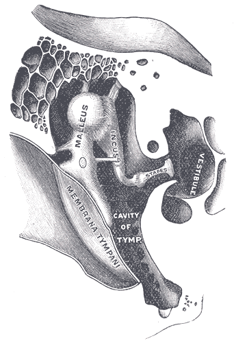
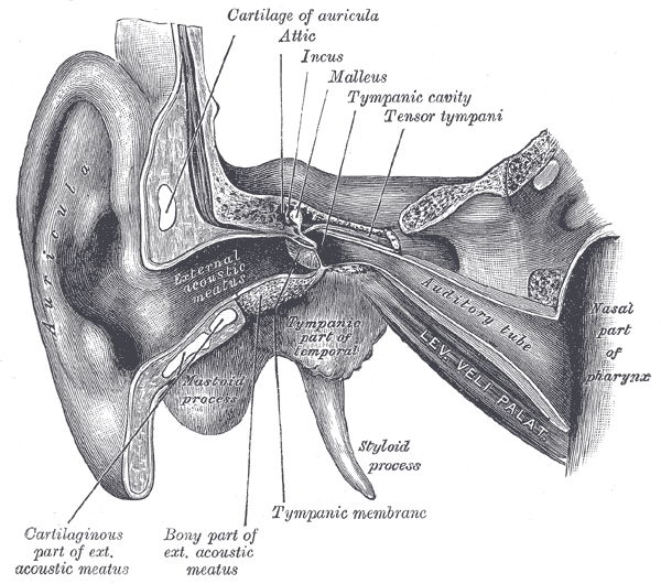
The middle ear (tympanic cavity or tympanum) is an air-filled box-shaped chamber with concave sides in the petrous part of the temporal bone. It is separated from the external ear by the tympanic membrane and from the inner ear by the medial wall. It contains the ossicular chain which functions to transmits and amplify sound vibrations from the tympanic membrane to the oval window of the lateral wall of the inner ear.
Structure
The middle ear is subdivided into several parts defined in relation to the planes of the tympanic membrane:
- Mesotympanum – directly medial to the membrane
- Epitympanum (attic, epitympanic recess) – superior to the membrane
- Hypotympanum – inferior to the membrane
- Protympanum – anterior to the membrane
- Retrotympanum – posterior to the membrane
Boundaries
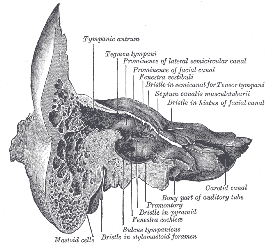
- Tegmental wall (roof) – formed by the tegmen tympani, a thin plate of petrous bone that also covers the canal for the tensor tympani muscle and the tympanic antrum. It separates the dura mater on the floor of the middle cranial fossa superiorly from the tympanic cavity.
- Jugular wall (floor) – formed by the jugular plate that separates the tympanic cavity from the superior bulb of the internal jugular vein.
- Membranous (lateral) wall – formed by the peaked convexity of the tympanic membrane and superiorly by the lateral bony wall of the epitympanic recess.
- Labyrinthine (medial) wall – separates the tympanic cavity from the inner ear. The initial part (basal turn) of the cochlea forms the promontory of the labyrinthine wall. It also features oval and round windows, which communicate with the inner ear.
- Mastoid (posterior) wall: This features the aditus (opening) to the mastoid antrum in its superior part, which connects the tympanic cavity to the mastoid cells. The canal for the facial nerve descends between the posterior wall and the antrum, medial to the aditus. Above this canal is the convex bulge of the lateral semicircular canal. Below the aditus there is a hollow conical projection, called the pyramid, which is perforated by the tendon of the stapedius muscle.
- Carotid (anterior) wall: This separates the tympanic membrane from the carotid canal. Inferiorly, there is the opening of the Eustachian tube (also known as the pharyngotympanic or auditory tube) and the semicanal for the tensor tympani muscle.
Bones
Middle ear ossicles consist of three small bones (the malleus, incus and stapes), which form a mobile chain across the tympanic cavity from the tympanic membrane to the oval window.
Malleus
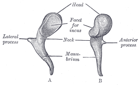
- Divided into four parts: the head, neck, short process, and handle
- The handle of the malleus attaches loosely to the tympanic membrane
- The head of the malleus attaches to the incus and forms the incudo-malleolar joint
Incus
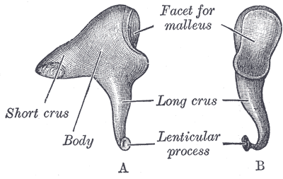
- Divided into the body, short process, and long process
- The incus connects with the stapes and forms the incudo-stapedial joint
Stapes
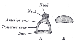
- Consists of the head, legs, and footplate
- The smallest bone in the human body
- The stapes articulates with the oval window of the inner ear
Muscles
There are two muscles which act to dampen over-vibration from low-pitched sound waves:
- Tensor tympani muscle
- Inserts into the handle of the malleus
- Innervated by the medial pterygoid nerve (branch of the mandibular division of the trigeminal nerve)
- Stapedius muscle
- Inserts into the neck of the stapes
- The smallest skeletal muscle in the human body
- Innervated by nerve to stapedius, a branch of the facial nerve
Innervation
- Chorda tympani, a branch of nervus intermedius, leaves the facial nerve in the facial canal and enters the tympanic cavity through the posterior wall, lateral to the pyramid, lying just underneath the mucous membrane. It runs over the pars flaccida of the tympanic membrane, and the neck of the malleus. It leaves at the anterior margin of the tympanic notch.
Arterial Supply
- Anterior tympanic artery from the maxillary artery (external carotid artery)
- Stylomastoid artery from the posterior auricular or occipital arteries – branch of external carotid artery
- Numerous small vessels from the external carotid artery
Venous Drainage
- Drainage to the pterygoid venous plexus and the superior petrosal sinus
Lymphatic Drainage
Drainage is to the parotid, retropharyngeal and upper group of deep cervical nodes.
Innervation
- Tympanic plexus – formed by the tympanic branch of the glossopharyngeal nerve (Jacobson nerve) and the sympathetic fibers from the internal carotid nerve. It supplies sensory and vasomotor fibres to the mucous membrane of the tympanic cavity, tympanic membrane and external auditory meatus. It also gives off the lesser petrosal nerve, which provides parasympathetic innervation to the parotid gland via the otic ganglion.
- The middle and external ear are also supplied by branches of the trigeminal, facial, glossopharyngeal and vagus nerves which results in referred pain in the ear from other areas supplied by these nerves, e.g. the teeth, posterior part of the tongue, pharynx and larynx.
Embryology
- The three ossicles are derived from the neural crest of the first and second pharyngeal arches during the sixth week of development.
- The malleus and incus are predominantly derived from the first arch from a structure called Meckel’s cartilage.
- The stapes is formed from the second arch from a structure called Reichert’s cartilage.
- Outer portions of the stapes footplate are derived from the mesoderm.
- Throughout the fetal period, the ossicles fully ossify through a process of endochondral ossification. This process of ossification occurs initially with the incus at 16 weeks, malleus at 16-17 weeks, and the stapes at 18 weeks. Ossification continues for up to 26 weeks.
Anatomical Variants
- Oval stapes
- Round stapes
- Triangular stapes
- Tunnel shaped stapes
- Asymmetrical
- Variation in size