

Overview
The vagus nerve (‘wandering’ nerve) is the tenth and longest cranial nerve (CN X) and has mixed sensory, motor and parasympathetic function. It provides the bulk of the parasympathetic input to the gastrointestinal system and to the heart.
- Sensory – Innervates the skin of the external acoustic meatus and the internal surfaces of the laryngopharynx and larynx. Provides visceral sensation to the heart and abdominal viscera.
- Special Sensory – Provides taste sensation to the epiglottis and root of the tongue.
- Motor – Provides motor innervation to the majority of the muscles of the pharynx, soft palate and larynx.
- Parasympathetic – Innervates the smooth muscle of the trachea, bronchi and gastrointestinal tract and regulates heart rhythm.
Nuclei
There are four cranial nerve nuclei that contribute to the vagus nerves:
Motor
- Dorsal nucleus of vagus – parasympathetic motor fibres (visceral efferent) supply the gastrointestinal tract and lungs
- Nucleus ambiguus – a large motor neuron embedded in the reticular formation, which provides motor fibres to the glossopharyngeal nerve, vagus nerve and spinal accessory nerve. In the vagus nerve, it supplies the soft palate, pharnyx and larynx
Sensory
- Solitary nucleus – general sensory taste (visceral afferent)
- Afferent fibers from facial nerve, glossopharyngeal nerve and vagus nerve convey taste in its rostral portion, and general visceral sense (general visceral afferent fibers) in its caudal portion
- Spinal trigeminal nucleus – general sensory (somatic afferent)
Origin
- Emerge as rootlets from post-olivary sulcus of the medulla, between glossopharyngeal rootlets above and cranial root accessory below
Segments & Course
Cisternal segment
- Unites into a single nerve and runs laterally through the medullary cistern
- Exits cranial cavity through the middle compartment (pars vascularis) of the jugular foramen, accompanied by inferior petrosal sinus and glossopharyngeal nerve (anteriorly), accessory nerve and sigmoid sinus (posteriorly)
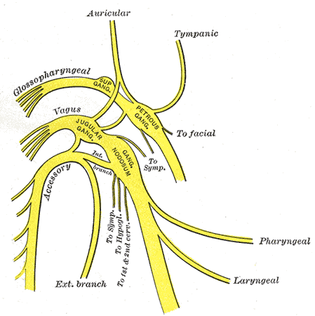
- Within the jugular fossa, beneath the skull base, lies two ganglia on the vagus nerve:
- Superior ganglion has cell bodies for the auricular branch – provides afferent general somatic innervation to the external ear and tympanic membrane
- Inferior ganglion has cell bodies of all the other sensory fibres in the vagus nerve – provides afferent general visceral fibres to the carotid and aortic bodies; the efferent fibres of this nerve travel to the nucleus tractus solitarius; the inferior ganglion also provides taste sensation to the pharynx and relays this information to the nucleus tractus solitarius
Cervical segment
- The fibres of the cranial root of the accessory nerve joins the vagus nerve above the inferior ganglion
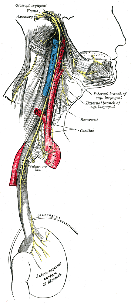
- Descends vertically in the neck within the carotid sheath, posterior and lateral to the internal carotid artery and common carotid artery and medial to the internal jugular vein

Thoracic segment
Left Vagus nerve
- Enters thorax via the thoracic inlet, between left subclavian artery (posteriorly) and vein (anteriorly)
- Descends in the superior mediastinum in a posterior direction to the left of the aortic arch, right of the left superior intercostal vein and behind left hilum
- The left vagus nerve travels anteriorly to the subclavian artery and enters the thoracic cavity wedged between the left common carotid and subclavian arteries
- It then descends posteriorly to the phrenic nerve and posterior to the left lung hilum, and travels medially towards to the oesophagus forming the oesophageal plexus with the right vagus nerve
Right Vagus nerve
- Enters thorax via the thoracic inlet, between right brachiocephalic vein and artery
- Descends in the superior mediastinum in a posterior direction, coming into contact with the right of the trachea posterior to the superior vena cava, left of azygous vein and posterior to the hilum
- Moves medially to contributes to the formation of the oesophageal plexus together with the left vagus nerve
Termination
- From the oesophageal plexus, the anterior and posterior vagal trunks emerge, to enter the abdomen through
the oesophageal hiatus in the diaphragm at the level of T10 and supplies the stomach- The anterior vagus gives hepatic branches
- The posterior vagus gives coeliac branches
- Through these branches, the vagus nerves supply the foregut, midgut, biliary tract and pancreas
Branches
From the superior ganglion:
- Small meningeal nerve – re-enters the skull at the jugular foramen, contains general afferent fibres and supplies the dura of the posterior cranial fossa
- Auricular nerve (Arnold’s nerve) – re-enters the lateral portion of the jugular foramen via the mastoid canaliculus. Contains general afferent fibres and innervates external tympanic membrane and small area of posterior external ear
- Small communicating branches
From the inferior ganglion:
- Pharyngeal nerve – supplies the pharyngeal plexus
- Supplies the superior, middle and inferior pharyngeal constrictor muscles, palatopharyngeus, salpingopharyngeus, levator veli palatini, palatoglossus and the muscle of the uvula
- Carotid body branches
- Superior laryngeal nerve which divides into the:
- Internal laryngeal nerve – enters the larynx through the thyrohoid membrane and supplies mucosa above the glottis
- External laryngeal nerve – travels to the inferior pharyngeal constrictor muscle. Innervates cricothyroid muscle
From the vagus nerve:
- Recurrent laryngeal nerve
- The left recurrent laryngeal nerve hooks beneath the aortic arch and around the ligamentum areriosum and ascends in the tracheoesophageal groove
- The right recurrent laryngeal nerve hooks beneath the subclavian artery and ascends in the tracheoesophageal groove to pass under the inferior constrictor of the pharynx behind the cricothyroid joint
- Superior and inferior cardiac nerves:
- Superior cardiac nerve – two branches (superior and inferior) on each side. Terminates in deep part of cardiac plexus.
- Inferior cardiac nerve – on the left arises from the recurrent laryngeal nerve. On the right arises form the trunk of the vagus. Both terminate in deep part of the cardiac plexus.
- Anterior and posterior bronchial nerves – locations refers to relation to root of the lung (i.e. anterior nerves located on anterior surface of the root of the lung). Innervates the bronchial tree and visceral pleura.
- Oesophageal plexus – formed by oesophageal branches of vagus nerve and visceral branches of the sympathetic trunk
- Gastric nerves – right vagus forms the posterior gastric plexus, left vagus forms the anterior gastric plexus.
- Coeliac plexus – coeliac branches primarily from the right vagus form the coeliac plexus, which innervates the pancreas, kidneys, spleen, adrenal glands and small bowel.
- Hepatic plexus – hepatic branches primarily from the left vagus form the hepatic plexus which innervates the liver
Blood supply
- The middle meningeal artery supplies the intracranial blood supply to the vagus nerve.
- The extracranial blood supply is from the common carotid artery, internal carotid artery, inferior thyroid artery, external carotid artery, a posterior meningeal artery, internal thoracic arteries, bronchial arteries, and esophageal arteries.
Lymphatics
The vagal system is involved in regulating the contraction of lymphatic (containing actin) cells.
Development
The vagus nerve arises from the fourth branchial arch
Variants
- The recurrent laryngeal nerve has two branches prior to inserting into the larynx; the branching is typically inferior to the cricoid cartilage; however, there are instances when there are more than two branches, and thus are called oesophageal branches.
- There are several variations of the non-recurrent vagus nerve.