

Overview
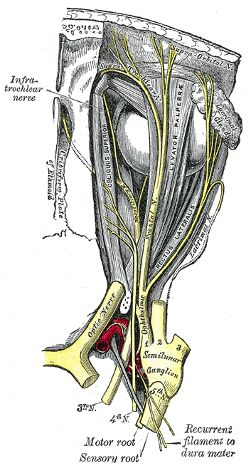
The oculomotor nerve is the third cranial nerve (CN III) and carries motor and parasympathetic innervation to structures within the bony orbit.
Function
The three main motor functions are:
- Innervation to the intrinsic eyes muscles controlling pupil diameter and lens shape (autonomic, parasympathetic)
- Innervation to the upper eyelid (somatic)
- Innervation of the extrinsic eye muscles that allow for visual tracking and gaze fixation (somatic) – except for superior oblique and lateral rectus muscle.
Origin
There are two nuclei from which the oculomotor nerve originates:
- The oculomotor nucleus at the level of the superior colliculus in the midbrain, ventral to the cerebral aqueduct, supplies somatic motor axons that innervate skeletal muscles associated with the eye
- The accessory oculomotor (Edinger-Westphal) nucleus supplies parasympathetic visceral motor fibres to muscles within the eye via the ciliary ganglion thus controls the sphincter pupillae (pupil constriction) and ciliary muscle (accomodation).

Course
- Originates from the oculomotor and Edinger-Westphal nucleus at the posteromedial aspect of the midbrain
- Fibres from both nuclei exit the anterior surface of the brainstem inferior to the mamillary bodies as the oculomotor nerve, invested with a sheath of pia mater and enclosed in a prolongation from the arachnoid.
- It then passes through the red nucleus, then via the substantia nigra exiting through the interpeduncular fossa
- It passes between the posterior cerebral artery (superiorly) and superior cerebellar (inferiorly) arteries.

- It then pierces the dura mater anterior and lateral to the posterior clinoid process covering the cavernous sinus, to traverse through the superolateral aspect of the cavernous sinus wall.
- After exiting the cavernous sinus, it enters the orbit via the superior orbital fissure then divides into a superior and inferior branch.
- Both branches traverse the orbit within the boundaries of the common tendinous ring (annulus of Zinn)
Divisions & Supply
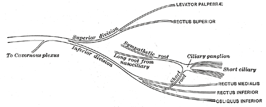
Superior Division
- Runs forward above the optic nerve
- Provides motor innervation to the:
- Superior rectus: elevation, adduction, medial rotation of the eyeball
- Levator palpebrae superioris: elevation of the upper eyelid
- Carries sympathetic fibres from the internal carotid-cavernous plexus to the smooth muscle of the superior tarsal muscle.
Inferior Division
- Larger than the superior division. Further divides into three branches to provide motor innervation to the:
- Inferior rectus: depression, adduction, lateral rotation of the eyeball
- Medial rectus: adduction of eyeball
- Inferior oblique: abduction, elevation, lateral rotation of the eyeball
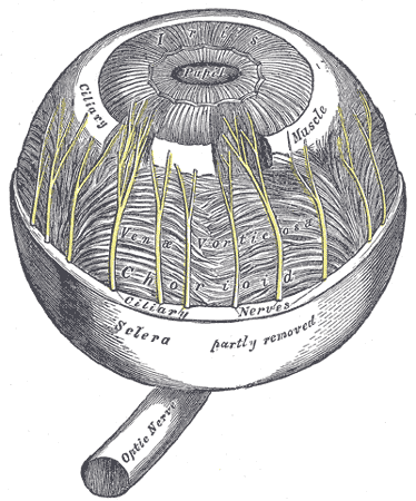
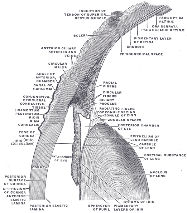
- Supplies pre-ganglionic parasympathetic fibres to the ciliary ganglion, which innervates the intrinsic muscles of the eye:
- Sphincter pupillae – constricts the pupil
- Ciliary muscles – changes shape of the lens for accomodation of light
Blood supply
- The vasa vasorum supplies the inner somatic (voluntary) nerve fibres while pia mater blood vessels supply the outer autonomic nerve fibers
- Both the oculomotor nucleus and Edinger-Westphal nucleus are both located in the medial midbrain, which is supplied by the paramedian branches of the upper basilar artery and the proximal posterior cerebral artery.
Development
Derived from the basal plate of the embryonic midbrain
Relations
- The trochlear nerves and ophthalmic and maxillary divisions of CN5 lie below the 3rd nerves in the lateral dural wall of the cavernous sinus.
- The abducens nerves course within the cavernous sinus itself.
Imaging Appearance
General
- Intercavenourse course is best appreciated on coronal plane.
- Superior most-aspect of the lateral dural wall of the cavernous sinus
MRI
- High-resolution coronal T2: A thin bright rim of CSF, representing the oculomotor cistern, surrounds the nerves for a variable distance within the cavernous sinus.