

RANZCR Anatomy Syllabus
Category 1
• Boundaries & walls, including contributions from specific skull bones
• Foramina & contents
• Optic canal
• Superior orbital fissure
• Inferior orbital fissure
Category 2
• Lacrimal fossa/crest
• Periorbita
• Medial & lateral tubercles
• Orbital septum
• Boundaries & walls, including contributions from specific skull bones
• Foramina & contents
• Optic canal
• Superior orbital fissure
• Inferior orbital fissure
Category 2
• Lacrimal fossa/crest
• Periorbita
• Medial & lateral tubercles
• Orbital septum
Bony Orbit |
|---|
| CATEGORY 1SEP 2013 | APR 2016 |
Gross Anatomy
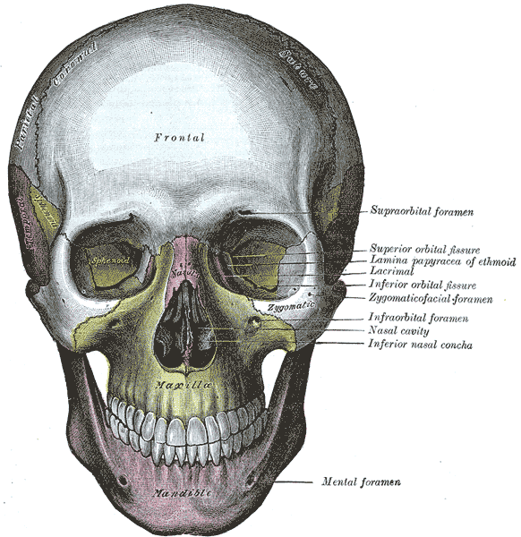
- paired pyramidal bony cavities invaginating the skull in the mid-face region and enclosing the eyeball.
- composed of 7 bones forming four-walled pyramid where apex is pointing posteriorly and base is situated anteriorly.
Relations
- superiorly: anterior cranial fossa.
- medially: nasal cavity and ethmoid sinus.
- inferiorly: maxillary sinus.
- posterolateral: infratemporal fossa and middle cranial fossa.
The walls of the bony orbit are invested by the periorbita, a periosteum continuous with that of the facial bones and dura mater (covering the optic nerve) posteriorly.
Boundaries
Roof (superior wall)
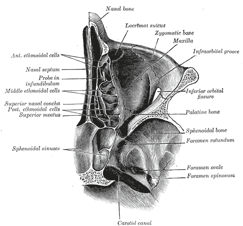
- formed by two bones: the orbital part of the frontal bone and lesser wing of the sphenoid posteriorly.
Floor (inferior wall)
- formed by three bones;
- the orbital surface of the maxilla (grooved and canalised by the infraorbital nerve)
- zygomatic bone laterally
- orbital process of the palatine bone posteriorly
Medial wall
- Formed by four bones: the ethmoid, maxilla, lacrimal and the lesser wings sphenoid bones.
- The medial walls of both orbits are parallel to each other.
- Lamina papyracea: the paper-thin aspect of the medial orbital wall.
- The ethmoid bone separates the orbit from the ethmoid sinus.
Lateral wall
- formed by two bones: the zygomatic bone (thickest and strong part of the orbital walls) and greater wing of the sphenoid.
- the lateral walls of both orbits meet at 90 degrees.
Apex
- Located at the opening to the optic canal, the optic foramen.
Base
- Opens out into the face, and is bounded by the eyelids
Variants
- Presence of middle ethmoid foramen
- Haller air cells
- Onodi cells
- Pneumatised lesser wing of sphenoid
- Supraorbital air cells
Superior Orbital Fissure |
|---|
| CATEGORY 1SEP 2013 | APR 2016 |
Shape, size, location
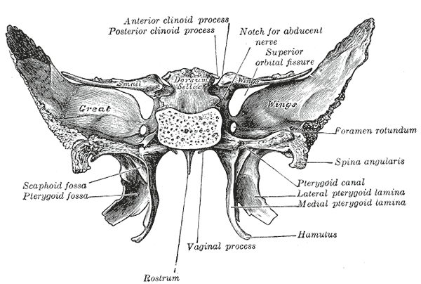
- Lies between the caudal surface of the lesser wing and medial border of the greater wing of the sphenoid bone.
- Communication between the cavernous sinus and apex of the orbit.
Boundaries
- Medial: body of sphenoid
- Superior: lesser wing of sphenoid
- Inferior: greater wing of sphenoid
- Lateral: frontal bone
Contents
Contents outside the Annulus of Zinn (superior to inferior):
- Lacrimal and frontal branches of ophthalmic nerve (CN VI)
- Superior ophthalmic vein
- Trochlear nerve (CN IV)
- Superior branch of inferior ophthalmic vein
Contents within the Annulus of Zinn:
- Superior and inferior divisions of oculomotor nerve (CN III)
- Abducens nerve (CN VI)
- Nasociliary branch of ophthalmic (CN VI)
(Note: The ophthalmic artery is transmitted with the optic nerve through the optic canal, which also is contained within the Annulus of Zinn but does not form part of the superior orbital fissure)
Relations
- Medially – the sphenoid sinus, optic canal (superomedial)
- Inferiorly – inferior orbital fissure, orbitalis muscle fibres, rotundum (inferolateral)
- Anteriorly – the extraconal orbit, with fat and ocular muscles/nerves
- The common tendinous ring straddles the superior orbital fissure.
- Superiorly – above the tendinous ring, the levator palpebrae superioris and superior oblique muscles take origin.
Inferior Orbital Fissure |
|---|
| CATEGORY 1NOT EXAMINED |
Opens into posterolateral aspect of orbital floor. It joins medially with the pterygomaxillary fissure at a right angle.
Boundaries
- Superiorly: greater wing of sphenoid
- Inferiorly: maxilla and orbital process of palatine bone
- Laterally: zygomatic bone.
Contents
Superior to inferior:
- Inferior branch of inferior ophthalmic vein
- Orbital ganglionic branches of the pterygopalatine ganglion from the maxillary division of the trigeminal nerve
- Infra-orbital nerve (CN Vb) – branch of the maxillary division of trigeminal nerve
- Infraorbital artery – branch of maxillary artery
- Infraorbital vein – branch of maxillary vein
- Zygomatic nerve (CN Vb) – branch of the maxillary division of trigeminal nerve