

RANZCR Syllabus
Category 1
• Ethmoid bone
• Frontal bone & sinus
• Sphenoid: Lesser wing
• Olfactory bulb and tract
Category 2
• Meningeal coverings
• Crista galli
Category 3
Nil
• Ethmoid bone
• Frontal bone & sinus
• Sphenoid: Lesser wing
• Olfactory bulb and tract
Category 2
• Meningeal coverings
• Crista galli
Category 3
Nil
General Description
The anterior cranial fossa is a depression in the floor of the cranial base which lodges the frontal lobes of the cerebral hemispheres. It is formed by the frontal, ethmoid and sphenoid bones.
Structures
Structures present in the midline of the anterior cranial fossa from anterior to posterior are:
- groove for superior sagittal sinus
- groove for anterior meningeal vessels
- foramen cecum – may be impervious. If open, transmits the emissary vein to the superior sagittal sinus
- crista galli – attachment for the falx cerebri
- slit and groove for nasociliary nerve
- anterior ethmoid foramen – transmits the anterior ethmoidal artery, nerve and vein
- orifices for olfactory nerve
- posterior ethmoid foramen – transmits the posterior ethmoidal nerve and vessels.
- olfactory fossa
Boundaries
- anterior: the inner surface of the frontal bone
- posterior: sharp posterior edge of the lesser wings of the sphenoid bone
- lateral floor: ridged orbital plates of the frontal bone
- medial floor: cribriform plate of the ethmoid bone
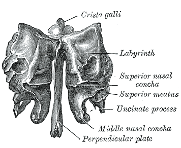
Shape, size, location
- Small unpaired bone, spongy structure
- Midline structure of the anterior cranium
- Forms roof of the nasal cavity and nasal septum
- Forms medial wall of the orbit
Structure
- Cribriform plate – a perforated plate of the ethmoid bone
- Perpendicular plate – forms superior two-thirds of the nasal septum
- Ethmoidal labyrinth – pair structured either side of the perpendicular plate, large masses containing the ethmoidal air cells
- Lamina papyracea (orbital plate of the ethmoid bone) – paper-thin, smooth, oblong bony plate located on the lateral surface of the labyrinth.
- Superior nasal concha and meatus
- Middle nasal concha
- Uncinate process – sickled-shaped projecting posteroinferiorly
Articulations
- Paired – nasal bones, maxillae, lacrimal bones, orbital process of palatine bones, inferior conchae.
- Unpaired – orbital plate of the frontal, vomer and sphenoid
Clinical significance
- Fracture of the lamina papyracea (medial wall of the orbit) can cause entrapment of the medial rectus muscle of the eye.
Shape, size, location
- The adult frontal bone is an unpaired bone forming the anterior and superior portions of the skull
Articulations
- Externally
- Anterior-medial: nasal bone (forming the nasion)
- Anterior-lateral: superior aspect of maxillary bone bilaterally.
- Lateral from maxillary bone: various bones that form posterior aspect of orbit
- Lacrimal bone, sphenoid bone, zygomatic bone.
- Posterior and lateral to the zygomatic bones: temporal surface of greater wing of sphenoid.
- Posteriorly: parietal bones.
- Internally
- Frontal bone connects with the ethmoid bone inferiorly and medially.
- Posteriorly and inferiorly, the frontal bone meets the sphenoidal bone.
- Posteriorly and laterally: temporal bone.
- Superior: parietal bone.
Landmarks
- Bregma: where sagittal and coronal sutures meet, represents position of anterior fontanelle in the young child.
- Pterion: joining of frontal, sphenoidal, parietal, and temporal
- Sinuses: anterior aspect of frontal skull contains two sinuses each superior to the orbital roof
Shape, size & location