

Overview
The cavernous sinus is a paired dural venous sinus, located bilaterally to the sella turcica and body of the sphenoid bone between the endosteal and meningeal layers of the dura. The left and right cavernous sinus communicate with each other by intercavernous sinuses anterior and posterior to the infundibulum of the pituitary gland. Each sinus is divided into numerous fibrous septa or ‘caves’ (hence cavernous) and is the only vein in the body to truly encapsulate an artery. Its function is to facilitate heat exchange between arterial and venous blood as they traverse each other.

Contents
Contents within the lateral wall:
- Exits the midbrain ventrally at the interpeduncular fossa and pierces the dura
- Enters the cavernous sinus, where it runs on the roof and lateral wall.
- After exiting the cavernous sinus, it enters the superior orbital fossa where it splits into the superior and inferior division.
- Originates from the trochlear nerve nucleus; it crosses the midline and emerges inferior to the inferior colliculus, situated in the posterior part of the midbrain.
- It then travels anteriorly around the midbrain, pierces and enters the dura mater near the tentorium cerebelli, and continues its course in the lateral wall of the cavernous sinus.
- After exiting the cavernous sinus, it enters the orbit through the superior orbital fissure to innervate the superior oblique muscle.
Ophthalmic nerve (CN V1)
- Exits the brainstem ventrally at the pontomedullary junction, pierces the dura, and travels the longest intracranial distance of all the cranial nerves
- CN V1 Branches of the trigeminal ganglion pass through the inferior part of the cavernous sinus
- After exiting the cavernous sinus, they enter the orbit via the superior orbital fissure.
Maxillary nerve (CN V2)
- External but immediately adjacent to the lateral wall
- CN V2 branches of the trigeminal ganglion enter the lower portion cavernous sinus and exit the skull via the foramen rotundum
Contents within cavernous sinus:
Abducens nerve (CN VI)
- Enters the back of the sinus after passing over the apex of the petrous temporal bone
- Lies inferolateral to the ICA
- Enters the superior orbital fissure at the anterior end of the sinus
C4 (cavernous) segment of the internal carotid artery
- Gives off meningohypophyseal trunk and inferolateral trunk as branches

Boundaries
- Medial wall – endosteum overlying the body of the sphenoid bone and pituitary gland/fossa
- Lateral wall – dura mater from the ridge of the roof to the floor of the middle cranial fossa
- Roof – fold of dura mater (meningeal and endosteal layers) attached to the anterior and middle clinoid processes
- Floor – endosteum overlying the base of the greater wing of the sphenoid bone
- Anterior wall – medial end of superior orbital fissure
- Posterior wall – petrous apex
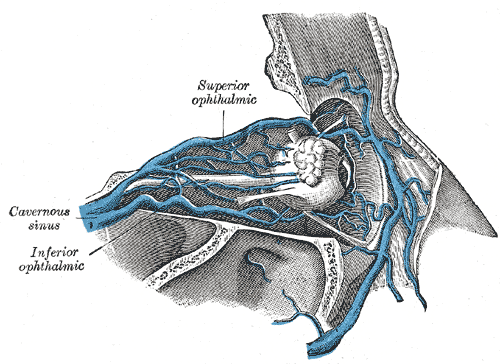
Tributaries
- Superior ophthalmic vein – Drains ethmoidal, nasofrontal, vorticose (drains ocular choroid) and central retinal veins
- Inferior ophthalmic vein – Drains eyelids, lacrimal sac, anterior floor and medial wall of the orbit
- Superficial middle cerebral vein (Sylvian vein) – Drains cerebral cortex
- Middle meningeal vein
- Hypophyseal veins
- Intercavernous sinuses
- Sphenoparietal sinuses
- Emissary veins
Drainage
Venous blood drains posteroinferiorly to eventually empty into the pterygoid plexuses
- Superior petrosal sinus to transverse sinus
- Inferior petrosal sinus directly to jugular bulb
- Venous plexus on the ICA to the clival basilar venous plexuses
- Emissary veins passing through the sphenoidal foramen, foramen ovale, foramen lacerum
Relations
- Superiorly: optic tract, optic chiasm, internal carotid artery
- Inferiorly: Foramen lacerum and the junction of the body and greater wing of sphenoid bone
- Medially: Pituitary gland and sphenoidal sinus
- Laterally: uncus of temporal lobe, trigeminal (Meckel’s) cave containing the trigeminal ganglion
- Anteriorly: superior orbital fissure and the apex of the orbit
- Posteriorly: apex of petrous temporal bone