

• Sella turcica
• Cavernous sinus, walls and contents
• Neurohypophysis & Stalk
• Adenohypophysis
• Pituitary blood supply & portal system
• Planum sphenoidale
Category 2
• Diaphragma sellae
Category 3
• ICA dural rings
Sella Turcica |
|---|
| CATEGORY 1PENDING |
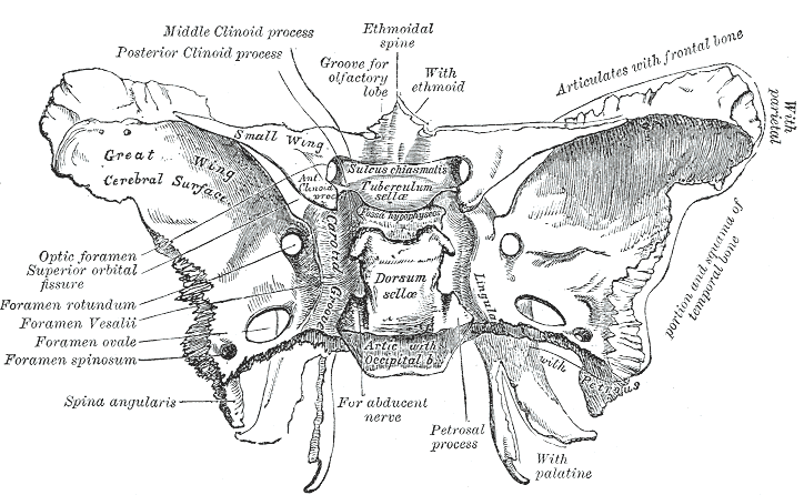
The sella turcica is a midline, dura-lined saddle-shaped depression in the upper surface of the sphenoid bone body, housing the pituitary gland.

Boundaries & Relations
- Anterior wall: tuberculum sellae, anterior clinoid processes
- ridged process of the sphenoid bone anterior boundary of the sella turcica
- Laterally: cavernous sinuses
- Superiorly (roof): diaphragma sella
- The sella diaphragm is a flat piece of dura mater with a circular fenestration allowing the vertical passage of the infundibulum (pituitary stalk)
- Anteroinferiorly: sphenoid sinus
- Posterior Wall: dorsum sellae, pontine cistern, posterior clinoid process,
- The posterior clinoid process deepen the sella turcica and give attachment to the tentorium cerebelli
- Anterolaterally: anterior clinoid process
- The anterior clinoid processes are formed by the medial and anterior projections of the lesser wing of the sphenoid bone
Cavernous Sinus
[copy_article id=”7406″]
Pituitary Gland |
|---|
| CATEGORY 1PENDING |
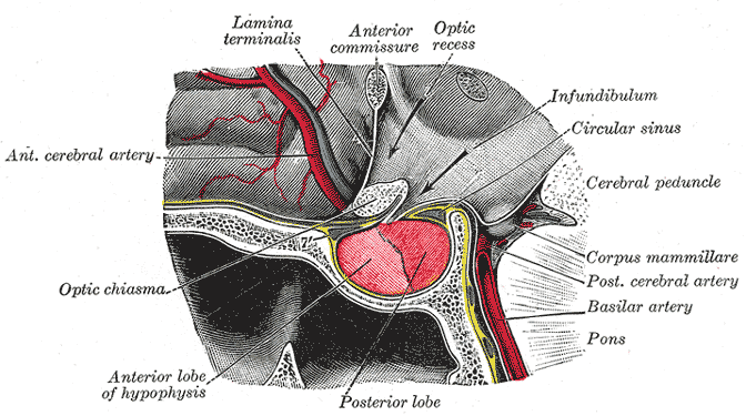
The pituitary gland (hypophysis cerebri) is a pea-sized oval endocrine gland protruding from the bottom of the hypothalamus, housed within the sella turcica of the sphenoid bone.

The tuber cinereum and median eminence of the hypothalamus give origin to an infundibulum. The tubular infundibulum connects the pituitary gland to the brain.
Structure
The pituitary gland is composed of the anterior lobe, intermediate lobe and infundibulum stalk.
Anterior lobe (Adenohypophysis)
Arises from an invagination of the oral ectoderm (Rathke’s pouch)
- Pars Distalis
- Distal part of the gland, forming the major bulk of the anterior lobe
- Most of the hormones get secreted from this region (GH, ACTH, TSH, FSH, LH and PRL)
- Pars Tuberalis
- An upwards extension of the pars anterior, surrounding the anterolateral aspect of the infundibulum
- Pars Intermedia
- Lies between the pars distalis and the posterior pituitary gland
Posterior lobe (neurohypophysis)
Originates from the neuroectoderm and develops as an extension of the hypothalamus from the floor of the third ventricle. It consists of the pars nervosa and the infundibular stalk.
- Pars Nervosa
- A neuroendocrine structure
- Infundibulum (pituitary stalk)
- Bridges the hypothalamic and hypophyseal system
Blood supply
- Anterior lobe: superior hypophyseal artery
- Originates from the internal carotid artery or posterior communicating artery
- Posterior lobe: inferior and middle hypophyseal artery
- Originates from the meningohypophyseal trunk, a branch of the internal carotid artery
Boundaries
- Superior: the gland is covered by a reflection of the dura mater – the diaphragm sellae. This membrane has a central opening allowing the passage of the infundibulum.
Relations
- Anteriorly: sphenoid sinus, anterior intercavernous sinus, tuberculum sellae, anterior clinoid process
- Posteriorly: posterior intercavernosus sinus, dorsum sellae, basilar artery, posterior clinoid process and pons
- Superiorly: diaphragma sellae, optic chiasm, suprasellar cistern, hypothalamus
- Inferior: sphenoid sinus
- Laterally: Cavernous sinus
Variants
- Hyperplasia: physiologic PRL cell hyperplasia due to lactation or pregnancy and in response to end organ failure such as Addison disease, Klinefelter, or Turner syndrome, or primary hypothyroidism
- Lateral flattening due to pressure of the carotid arteries
- Duplication: “empty sella sign”: occurs when diaphragm sellae is incomplete (normal variant) and CSF enters the pituitary fossa
- Rathke cleft cysts: arises from pars intermedia