

RANZCR Syllabus
Category 1
• Internal carotid arteries, branches & segments
• Ophthalmic artery and branches
• Circle of Willis configuration and common variations
• Middle cerebral artery (MCA), segments & branches
• Anterior cerebral artery (ACA), segments & branches
• Anterior communicating artery (AComA)
• Posterior cerebral artery (PCA), segments & branches
• Vertebral & basilar artery
• Anterior & posterior spinal arteries
• Posterior communicating artery (PComA)
• Cerebellar arteries (SCA, AICA, PICA)
• Arterial territories on crosssectional imaging, variations
Category 2
• Extradural ICA branches
– Inferolateral trunk
– Menigohypophyseal trunk
– Artery of Vidian canal
Category 3
• Anterior choroidal artery
• Anterolateral and anteromedial perforating arteries including artery of Heubner
• Intracranial – extracranial anastomoses
– Ophthalmic/facial
– Inferolateral & maxillary
• Posterolateral perforating arteries
• Posteromedial perforating arteries
• Basilar and vertebral perforators
• Internal carotid arteries, branches & segments
• Ophthalmic artery and branches
• Circle of Willis configuration and common variations
• Middle cerebral artery (MCA), segments & branches
• Anterior cerebral artery (ACA), segments & branches
• Anterior communicating artery (AComA)
• Posterior cerebral artery (PCA), segments & branches
• Vertebral & basilar artery
• Anterior & posterior spinal arteries
• Posterior communicating artery (PComA)
• Cerebellar arteries (SCA, AICA, PICA)
• Arterial territories on crosssectional imaging, variations
Category 2
• Extradural ICA branches
– Inferolateral trunk
– Menigohypophyseal trunk
– Artery of Vidian canal
Category 3
• Anterior choroidal artery
• Anterolateral and anteromedial perforating arteries including artery of Heubner
• Intracranial – extracranial anastomoses
– Ophthalmic/facial
– Inferolateral & maxillary
• Posterolateral perforating arteries
• Posteromedial perforating arteries
• Basilar and vertebral perforators
Overview
The internal carotid artery (ICA) is a major branch of the common carotid artery, supplying the head and brain, located bilaterally in the neck.
Origin
- Arises at bifurcation common carotid artery at the level of the upper border of the thyroid cartilage (level of C4).
- Commences with a slight bulge, the carotid sinus (containing baroreceptors supplied by glossopharyngeal and vagus nerves).
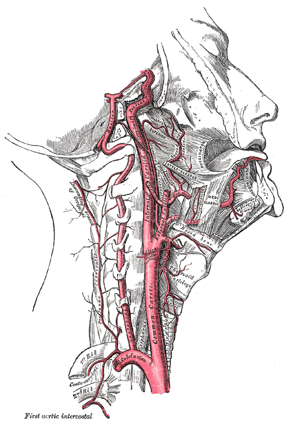
Segments & Course
C1 – Cervical
- Commences from common carotid bifurcation, the ICA ascends vertically within the carotid sheath – anterior to the transverse process of C1-C3
- Enter the base of skull via the carotid canal of the petrous temporal bone
C2 – Petrous
- Bends anteromedially then passes horizontally to the foramen lacerum
C3 – Lacerum
- From the foramen lacerum (does not pass through), curves upwards to enter the cavernous sinus through the petrolingual ligament
C4 – Cavernous
- Commences at the petrolingual ligament and has a curve in its course within the cavernous sinus (carotid siphon)
- Extends to the proximal dural ring (formed by medial and inferior periosteum of the anterior clinoid process)
C5 – Clinoid
- From proximal dural ring to the distal dural ring
C6 – Ophthalmic
- From distal dural ring, courses horizontally and parallel to the optic nerve, to the origin of posterior communicating artery
C7 – Communicating
- Passes between the optic and oculomotor nerves to the anterior perforated substance at the medial extremity of the lateral cerebral fissure before terminating as the anterior cerebral and middle cerebral artery
Termination
Anterior cerebral and middle cerebral artery
Branches
- C1: Cervical segment – none
- C2: Petrous (horizontal) segment
- Caroticotympanic artery
- Vidian artery
- C3: Lacerum segment – none
- C4: cavernous segment
- Meningohypophyseal trunk
- Inferolateral trunk
- Capsular arteries (of McConnell) (variable)
- C5: Clinoid segment – none
- C6: Ophthalmic (supraclinoid) segment
- Ophthalmic artery
- Superior hypophyseal artery
- C7: communicating segment
- Posterior communicating artery
- Anterior choroidal artery
- Anterior cerebral artery
- Middle cerebral artery
Arterial Supply
- Vasovasorum – small network of capillaries supplying the lumen and walls of large blood vessels
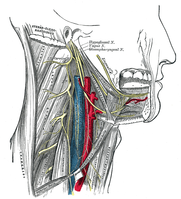
Relations
- Lateral to external carotid at its origin, but ascends more posteriorly, medially and deeper within the carotid sheath (beside the pharynx) to the carotid canal in the base of the skull.
- Anterior to sympathetic trunk, pharyngeal veins and superior laryngeal branch of the vagus nerve
- Lateral to ascending pharyngeal artery
- Medial to internal jugular vein
C1
- Posterior: longus capitis, superior cervical ganglion of the sympathetic trunk, superior laryngeal branch of vagus nerve
- Medial: pharynx and ascending pharyngeal artery
- Lateral: internal jugular vein and vagus nerve
- Anterior:
- Inferior: lingual vein, facial vein, occipital artery, and hypoglossal nerve
- Superior: separate from ECA by styloglossus, stylopharyngeus, tip of styloid process and stylohyoid ligament, glossopharyngeal nerve and pharyngeal branch of vagus nerve
Variant Anatomy
- Aberrant ICA (enlargement of caroticotympanic/hyoid artery which anastomoses with inferior tympanic artery from ECA, when cervical ICA is hypoplastic/absent)
- Absent ICA
- Retropharyngeal ICA
- Kissing carotids
- Persistent carotid-vertebrobasilar anastomsoes
- Lateralised ICA
Clinical significance
Surgically accessed via incision along anterior border of sternocleidomastoid.
A single artery supplying part of the posterior circulation of the brain.