

Overview
The vertebral arteries are major paired arteries of the neck that supply the vertebrobasilar vascular system.
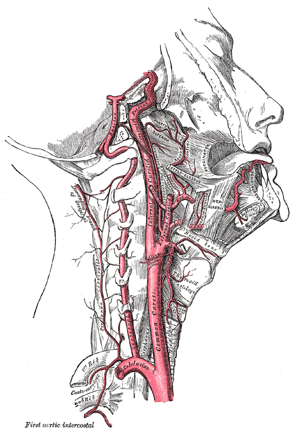
Origin
Arises from the posterosuperior part of the subclavian arteries bilaterally, although the origin can be variable.
Segments & Course
V1 (pre-foraminal/extra-osseous) segment
- From the origin, V1 courses posteriorly to pass between longus colli medially and anterior scalene laterally, through the colliscalene triangle
- It then enters the transverse foramen of C6
- anteriorly: common carotid artery, vertebral vein, internal jugular vein, thoracic duct (left VA), and lymphatic duct (right VA)
- posteriorly: ventral rami of spinal nerves C7 and C8, transverse process of C7, inferior cervical ganglion, sympathetic trunk
- anteromedially: inferior thyroid artery, middle cervical ganglion
V2 (foraminal) segment
- From the transverse foramen of C6, V2 ascends through the transverse foramina of the cervical vertebrae accompanied by the vertebral veins/plexi and the sympathetic nerves
- Posteriorly: Trunks of the cervical spinal nerves
V3 (atlantic, extradural or extraspinal) segment
V3 is tortuous, providing length and freedom for the vessel to stretch during rotation of the head at the atlantoaxial joints.
- After passing through the transverse foramen of C2, V3 sweeps laterally over its transverse process.
- It then curves superiorly passing immediately lateral to the lateral mass of C2 and the lateral C1/2 articulation before passing through the transverse foramen of C1.
- Exiting this foramen, V3 then once again courses posteriorly around the posterolateral border of the lateral mass of C1. Passing superomedially it grooves the upper surface of the posterior arch of C1
- It then enters the vertebral canal by piercing the posterior atlantooccipital membrane, spinal dura and arachnoid mater to continue as V4.
V4 (intradural or intracranial) segment
- From the dura at the lateral edge of the posterior atlantooccipital membrane,
- V4 ascends anterior to the roots of the hypoglossal nerve (CN XII) and joins its contralateral counterpart at the pontomedullary junction to form the basilar artery.
Termination
- Basilar artery at the pontomedullary junction.
Branches & Supply

Anterior spinal artery
- Arises within the intradural segment as two small vessels from each vertebral artery that unite around the spinal cord
- Then passes through the foramen magnum, descending along the anterior aspect of the spinal cord
- Supplies the upper cervical spinal cord, inferior medulla
Posterior spinal artery
- May arise from the posterior inferior cerebellar artery
- Supplies dorsal spinal cord to conus medullaris
Posterior inferior cerebellar artery
- Largest branch of the vertebral artery
- Originates from each of vertebral arteries intracranial segment and supplies the cerebellum
- Supplies lateral medulla, tonsil, inferior vermis/cerebellum, choroid plexus of 4th ventricle
Meningeal branches
- From the intracranial segment, near the foramen magnum
- Supplies the meninges
Medullary arteries
- From the intracranial segment
- Supplies medulla oblongata
Variant anatomy
- asymmetry due to vertebral arterial hypoplasia, absence or termination into PICA of one of the vertebral arteries is very common
- left dominant ~45% (range 42-50%)
- right dominant ~30% (range 25-32%)
- co-dominant ~25% (range 25-26%)
- complete or partial vertebral artery duplication
- vertebral artery fenestration (predisposes to aneurysms)
- variable origin
- single left aberrant origin (86%), single right aberrant (12%), bilateral aberrant origin (3%)
- aortic arch origin of left vertebral artery: incidence ~5% (range 3.1-8.3%)
- second (not first) branch of subclavian artery
- external carotid artery (rare)
- common carotid artery (rare)
- internal carotid artery (rare)
- ostium may have variable orientation
- cranial ~47%
- posterior ~45%
- caudal ~5%
- anterior ~3%