

Overview
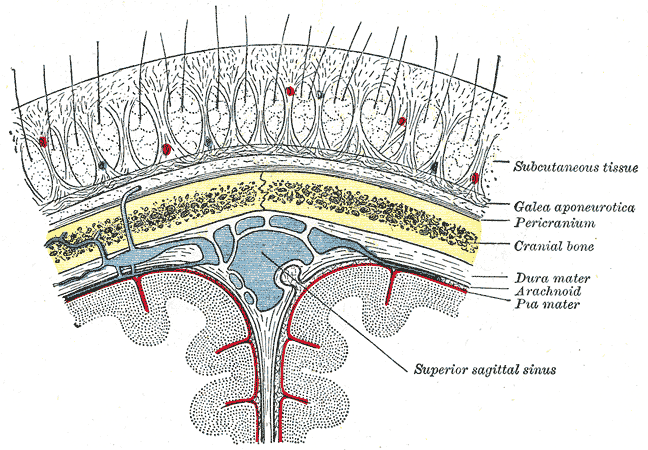
The meninges are membranous coverings of the brain and spinal cord. The dura mater is the outermost layer of meninges composed of two layers (lamellae), the periosteal (endosteal) layer and the meningeal layer. The general functions include:
- Envelop and protect the brain and spinal cord
- Regulate the generation and migration of neural progenitors
- Limit the rotational displacement of the brain
- Regulate survival and proliferation of radial glial
- Regulate axon behaviour at CNS PNS interface
Structure
The periosteal and meningeal layers are firmly adherent except when separated to enclose the dural venous sinuses, including the cavernous sinus.
Periosteal (endosteal) layer
- Outer layer lining the inner surface of the inner calvarium, essentially the periosteum.
- As the periosteum surrounds the cranial bones, the dura mater, therefore, extends into the skull sutures and foramina
- This explains why epidural haematomas do not cross suture lines
- It does not extend beyond the foramen magnum to become contiguous with the dura mater (i.e. there is no endosteal layer of the spinal cord).
- It also provides tubular sheath-like covering for the cranial nerves and arteries. As cranial nerves and vessels exit the skull, the endosteal layer fuses with the epineurium of nerves and the tunica adventitia of arteries such as the carotid and vertebral arteries
Meningeal layer
- Inner layer
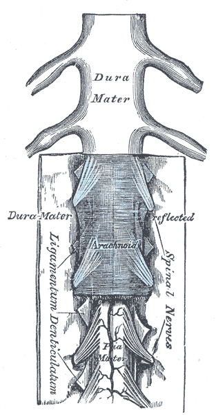
- Durable, dense, fibrous membrane continuous with the dura mater of the spinal cord via the foramen magnum. When it covers the spinal cord it may be known as the dural or thecal sac.
- Creates several inward folds to form dural reflections
The two dural layers are firmly attached except in places where they separate to enclose the dural venous sinuses.
– Inner meningeal layer folds inward forming the falx cerebri, falx cerebelli, tentorium cerebeli and the diaphragma sellae.
Also continues extracranially into the orbit and through the foramen magnum into the spinal canal. It fuses with the
epineurium of cranial/peripheral nerves and the adventitia of the carotid and vertebral arteries
– Tightly adherent to the skull at sutural attachments
Dural reflections
The dural mater folds inwards upon itself to form four dural reflections:

Falx cerebri
- Large, sickle-shaped meningeal projection of the dura located at the midline between the two cerebral hemispheres.
- Stiffer than surrounding tissue, thus dampens brain movement within the skull vault
Tentorium cerebelli
- Second largest, crescent-shaped fold forming a roof over the posterior cranial fossa, separating the occipital lobes from the cerebellum
Falx cerebelli
- Lies inferior to the tentorium cerebelli
- Vertical infolding separating the cerebellar hemisphere
Diaphragma sellae
- Small circular infolding forming the roof of the sella turcica, covering the pituitary gland and sella turcica
- The diaphragma sellae has a central perforation to allow passage of the pituitary gland stalk
Potential spaces
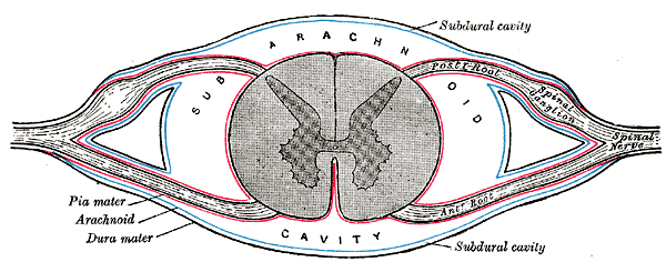
Cranial epidural (extradural) space
- Potential space between the skull and dura mater
- Contains meningeal arteries and veins
- Branches of the middle meningeal artery can be damaged from trauma, resulting in a contained collection of blood known as an epidural haematoma.
Spinal epidural space
- Space between the spinal dural mater (meningeal layer only) and the vertebral column – extends from the foramen magnum to the sacral hiatus
- Separate from and not continuous with the cranial epidural space
- Contains epidural fat, lymphatics, blood vessels, and nerve roots
Subdural space
- Formed by tearing the subdural compartment between neuroethelial cells alongside the collage fibres of the dura mater
Blood Supply
Anterior cranial fossa
- Anterior meningeal artery – a branch of the anterior ethmoidal artery
- Meningeal branches of the ophthalmic artery
Middle cranial fossa
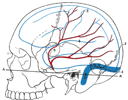
- Middle meningeal artery – dominant supply of the cranial dura. It branches off the maxillary artery enters via foramen spinosum
- Anterior division: runs anterolaterally on the greater wing of the sphenoid before coursing superiorly, often grooving the bone, and passes under the pterion before giving off terminal branches over the upper parietal bone
- Posterior division: runs horizontally posteriorly over the squamous part of the temporal bone to give rise to its terminal branches over the lower parietal bone
- Accessory meningeal arteries – branches from the maxillary artery
- Small meningeal branches of the internal carotid artery and accessory meningeal arteries – supply the dura over the cavernous sinus
Posterior cranial fossa
- Posterior meningeal artery – branch of the ascending pharyngeal artery
- Meningeal branch from the ascending pharyngeal artery, occipital artery and vertebral artery
Venous drainage
- Via multiple unnamed meningeal veins present in the periosteal layer, which follow branches of the middle meningeal artery and drain into the sphenopalatine sinus or pterygoid venous plexus
- The dural venous sinuses are between the periosteal and meningeal layers. These sinuses are responsible for the venous vasculature of the cranium. The sinuses converge and drain into the internal jugular vein.
Lymphatics
- Lymphatic drainage of the meninges is incompletely understood
Nerves
The dominant nerve supplying most of the supratentorial dura is the tentorial nerve (a branch of the ophthalmic nerve (CN Va) which supplies the falx cerebri, calvarial dura, and superior surface of the tentorium cerebelli.
Anterior cranial fossa
- Anterior meningeal branches from the ethmoidal nerves (CN Va)
- Meningeal branches from the maxillary nerve (CN Vb)
Middle cranial fossa
- Middle meningeal nerve which is a branch of the nerve (CN Vb) supplies the anterior half of the fossa
- Nervus spinosus which is the meningeal branch of the mandibular nerve (CN Vc) supplies the posterior half of the fossa
Posterior cranial fossa
- Sensory branches from the glossopharyngeal nerve (CN IX) and vagus nerve (CN X) supply the inferior surface of the tentorium and dura of the posterior fossa
- Small sensory branches of the first three cervical nerves (C1 to C3 dorsal rami) supply around the foramen magnum
Development
- The dural mater (pachymeninx) originates from somitic mesoderm, which condenses around the neural tube shortly after the neural tube closes at embryonic day 9
- The pia and arachnoid (leptomeninges) originate from the neural crest.
- The dura, forms between the arachnoid mater and the calvarial mesenchyme, which ultimately forms the calvarial bones.
- The first of the dural reflections to develop is the tentorium cerebelli.