

Overview
The glossopharyngeal nerve is the ninth paired cranial nerve (CN IX). It is one of the four cranial nerves that has sensory, motor, and parasympathetic functions:
- Sensory: Innervates the oropharynx, carotid body and sinus, posterior third of the tongue (general sensation), middle ear cavity and Eustachian tube.
- Generation sensation to the anterior two-thirds of the tongue is supplied by the lingual nerve, a branch of the mandibular division of trigeminal nerve.
- Special sensory: Provides taste sensation to the posterior third of the tongue
- Taste sensation to the anterior two-thirds of the tongue is supplied by the chorda tympani nerve, a branch of the facial nerve.
- Parasympathetic: Provides parasympathetic innervation to the parotid gland – produces secretion of saliva
- Motor: Innervates the stylopharyngeus muscle of the pharynx (a long, slender and tapered longitudinal muscle that has a functional role in swallowing)

Origin
There are four cranial nerve nuclei in the lower pons and medulla:
- Solitary nucleus – located in the dorsolateral medulla oblongata and lower pons, which conveys taste sensation from the posterior third of the tongue and vallate papillae
- Sensory nucleus of the trigeminal nerve – ocated in the caudal aspect of the pons, which conveys somatic sensation
- Nucleus ambiguus – located in the rostral pons, the motor fibres receives bilateral supranuclear innervation from the corticobulbar fibres and supplies the stylopharyngeus muscle.
- Inferior salivary nucleus – located in the dorsal pons, gives rise to autonomic parasympathetic fibres which are carried in the lesser petrosal nerve via the tympanic branch to the otic ganglion. The postganglionic fibres are distributed to the parotid gland via the auriculotemporal nerve.
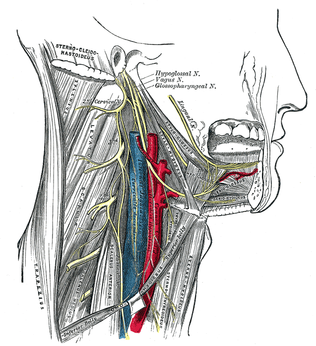
Course
- It emerges from the post-olivary sulcus of the medulla, traveling laterally across the flocculus in the posterior cranial fossa.
- The nerve leaves the cranium via the pars nervosa of the jugular foramen in a separate sheath of dura mater
- Immediately outside the jugular foramen lie two ganglia – superior (jugular) ganglion and inferior (petrous) ganglion which contain the cell bodies of the sensory fibres in the glossopharyngeal nerve.
- Tympanic nerve (Jacobson nerve) arises from the inferior ganglion. It has a mixed sensory and parasympathetic composition. It enters the tympanic cavity via the inferior tympanic canaliculus (Jacobson canal) and contributes to the tympanic plexus located on the cochlear promontory. The parasympathetic fibres leave the plexus as the lesser petrosal nerve.
- The glossopharyngeal nerve descends down the neck, between the internal jugular vein and internal carotid artery.
- At the inferior margin of the stylopharyngeus, several branches arise to provide motor innervation to the muscle It also gives rise to the carotid sinus nerve, which provides sensation to the carotid sinus and body.
- It then enters the pharynx by passing between the superior and middle pharyngeal constrictors.
- Within the pharynx, it terminates by dividing into several branches
- Lingual – innervates the posterior third of the tongue
- Tonsillar
- Pharyngeal – usually three, course medially and anastomose with branches of the vagus to form the pharyngreal plexus
Development
The glossopharyngeal nerve is associated with the derivatives of the third pharyngeal arch.