

Overview
The vestibulocochlear nerve is the eighth cranial nerve and consists of two parts. It conveys sensory impulses from the organs of hearing and balance to the inner ear.

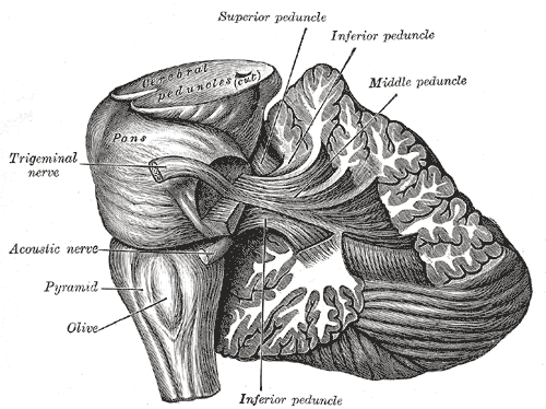
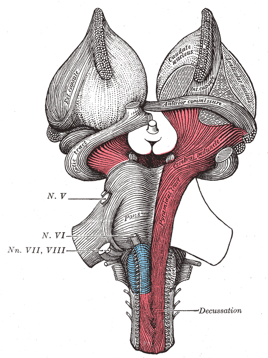
Origin
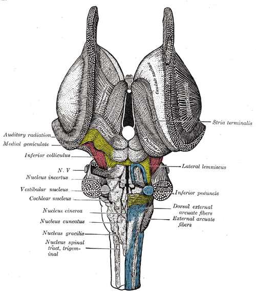
- Vestibular component – arises from four vestibular nuclei found laterally in the rhomboid fossa
- Superior vestibular (Bechterew’s) nucleus
- Lateral vestibular (Deiters’) nucleus
- Inferior vestibular (Roller’s) nucleus
- Medial vestibular (Schwalbe’s) nucleus
- Cochlear component – arises from two cochlear nuclei located medial to the inferior peduncle of the cerebellum in the lateral angle of the rhomboid fossa
- Posterior cochlear (dorsal) nucleus
- Anterior cochlear (ventral) nuclei
Course
- Both vestibular and cochlear fibres combine in the pons to form the vestibulocochlear nerve and emerge from the cerebellopontine angle within the posterior cranial fossa
- It travels anterolaterally to exit the cranium via the internal acoustic meatus of the temporal bone, coursing laterally to the abducens and facial nerve
- At the fundus of the internal acoustic meatus, the vestibulocochlear nerve splits to form the vestibular and cochlear nerve
- The cochlear nerve is located in the anterior inferior quadrant of the internal auditory canal, below the falciform crest (crista falicformis) and anterior to Bill bar.
- The vestibular nerve is located posterior to Bill bar and is divided into superior and inferior components by the falciform crest
- The falciform crest divides the internal auditory canal into superior and inferior compartments
- A vertical bony ridged named the Bill bar further divides the superior compartment into anterior and posterior compartments
- The nervus intermedius lies in between the facial nerve and the vestibulocochlear nerve – hence its name intermedius
- The vestibular nerve passes through the vestibular ganglion, then forms branches that innervate the vestibular system of the inner ear
- Utriculo-ampullary nerve
- Saccular nerve
- Posterior ampullary nerve
- The cochlear nerve penetrates the anterior lower quadrant of the fundus of the internal acoustic meatus and through the longitudinal canals of the modiolus, it reaches the spiral canal of the modiolus in which the spiral ganglion is placed. It innervates the cochlea of the inner ear.