

Overview
The facial nerve is the seventh cranial nerve. It controls muscles of facial expression and functions in taste sensation.
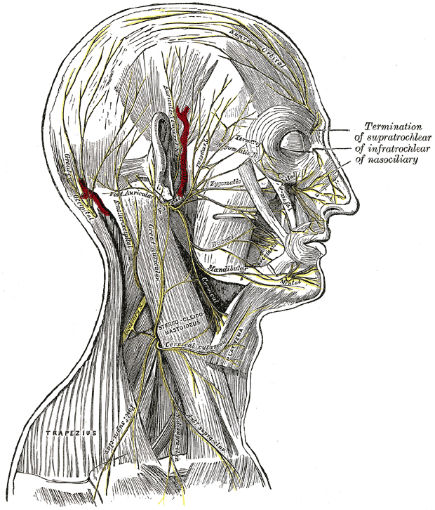
Functions
- Motor – Facial motor nucleus
- Muscles of facial expression (see below)
- Posterior belly of digastric (anterior belly – innervated by mylohyoid nerve, branch of the mandibular division of the trigeminal nerve)
- Stylohyoid
- Stapedius
- Special sensory – Solitary nucleus
- Chorda tympani – branch of facial nerve responsible for taste to anterior two-thirds of tongue
- Arises in facial canal, travels across bones of the middle ear, exits via the petrotympanic fissure, enters the infratemporal fossa, accompanies lingual nerve
- Chorda tympani – branch of facial nerve responsible for taste to anterior two-thirds of tongue
- Parasympathetic – Superior salivatory nucleus
- Submandibular gland, sublingual gland, lacrimal glands
Origin
- Arises from pontomedullary function as a large motor root and small sensory root (nervus intermedius)
Conveys:
- Afferent taste fibres from the chorda tympani – arise from the anterior two thirds of the tongue
- Taste fibres from the soft palate via the palatine and greater petrosal nerves
- Preganglionic parasympathetic innervation to the submandibular, sublingual and lacrimal glands

Segments, Branches & Course
Intracranial (cisternal) segment
- Motor root emerges at the lower border of the pons, above the olive, between the abducens nerve (medially) and the vestibulocochlear nerve (laterally)
- It is joined by nervus intermedius (of Wrisberg), which emerges lateral to the main trunk.
- Both travel laterally through the cerebellopontine angle to the internal acoustic meatus together with the vestibulocochlear nerve
- The nervus intermedius lies in between the facial nerve and the vestibulocochlear nerve – hence its name intermedius

Meatal (canalicular) segment
- Located in the anterior superior quadrant of the internal auditory canal, above the falciform crest (crista falicformis) and anterior to Bill bar.
- The falciform crest divides the internal auditory canal into superior and inferior compartments
- A vertical bony ridged named the Bill bar further divides the superior compartment into anterior and posterior compartments

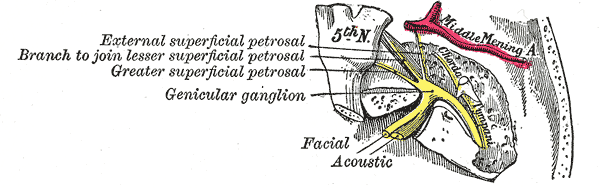
Labyrinthine (proximal) segment
- Facial nerve and nervus intermedius then enters the Fallopian canal of the petrous temporal bone
- The name of the segment is derived from the location of the segment immediately posterior to the cochlea. The nerve is posterolateral to the ampullated ends of the horizontal and superior semicircular canals and is anterior to the vestibule.
- It then bends posteriorly (the anterior or first genu) at the geniculate ganglion
- The geniculate ganglion is formed by the juncture of the nervus intermedius and facial nerve into a common trunk
- Labyrinthine segment is the shortest, narrowest and most susceptible to vascular compromise
- It also demonstrates normal contrast enhancement up to the stylomastoid foramen
Gives rise to:
- Greater petrosal nerve – carries preganglionic parasympathetic fibers to the lacrimal gland
- Nerve to the stapedius – motor fibres to stapedius muscle of the middle ear
Tympanic (horizontal) segment
- As the nerve passes posteriorly from the geniculate ganglion, it becomes the tympanic segment.
- The tympanic segment extends from the geniculate ganglion to the horizontal semicircular canal.
- It lies immediately beneath the lateral semicircular canal in the medial wall of the middle ear cavity
- The nerve passes posterior to the cochleariform process, tensor tympani and oval window.
- Distal to the pyramidal eminence the nerve turns vertically downwards
Gives rise to:
- No branches
Mastoid segment
- Extends from the posterior genu to the stylomastoid foramen, through the Fallopian canal
- It runs in the medial wall of the aditus ad antrum of the mastoid posterior to the facial recess of the mesotympanum
Gives rise to:
- Chorda tympani – transmits taste sensation from anterior two-thirds of the tongue
- Nerve to stapedius – supplies the stapedius muscle (attaches to the posterior aspect of stapes to dampen vibrations)
- Nerve from the auricular branch of the vagus nerve – carries pain fibres to the posterior part of the external acoustic meatus
Extratemporal segment
- As the nerve exits the stylomastoid foramen, it gives off the posterior auricular nerve that supplies part of the external acoustic meatus, tympanic membrane, auricular muscles, and occipital part of occipitofrontalis.
- It then passes between the posterior belly of the digastric muscle and the stylohyoid muscle whilst supplying both
- Anterior belly – innervated by mylohyoid nerve, branch of the mandibular division of the trigeminal nerve
- It then enters the parotid gland between the deep and superficial lobes.
- It divides into two main branches at the pes anserinus – superior temporofacial and inferior cervicofacial branches. From the anterior border of the gland, five branches emerge (from superior to inferior):
- Temporal – Frontalis, obicularis oculi, corrugator supercilii
- Zygomatic – Obicularis oculi
- Buccal – Obicularis oris, buccinator, zygomaticus muscles
- Marginal mandibular – Mentalis muscle
- Cervical branches – Platysma
Development
- By the third week of gestation, the facioacoustic primordium gives rise to the CN VII and VIII.
- During the fourth week, the chorda tympani can be discerned from the main branch
- By the fifth week, the geniculate ganglion, nervus intermedius, and greate petrosal nerve are visible
Imaging
- Principle of enhancement;
- Breakdown or absence of the blood–nerve barrier, or
- Presence of vascularised tissue
- Segments of the facial nerve within the CNS (intracranial) are protected by a tight blood–nerve barrier, like the blood–brain barrier.
- As the nerve exits the brainstem and traverses the temporal bone, parts of it lie outside this barrier, and may show physiological enhancement.
| Segment | Enhancement |
|---|---|
| Intracranial | No |
| Intracanalicular | Mild |
| Labyrinthine | Minimal |
| Geniculate ganglion | Yes |
| Tympanic | Faint |
| Mastoid | Faint |
| Extracranial | Variable |