

• Neocerebellum
• Vermis
• Cerebellar tonsils
• Dentate nuclei
• Superior, middle and inferior peduncles
Category 2
• Superior & inferior medullary velum
Category 3
Nil
Overview
The cerebellum, the largest part of the hindbrain (specifically the superior part termed the metencephalon), is located in the posterior cranial fossa. Its cortex is a thin (1mm), sheet-like structure, finely-folded like an accordion into horizontal parallel grooves termed folia fused at the midline. Its primary function is in the coordination of gait, maintenance of posture, control of muscle tone and voluntary muscle activity, however, it is unable to initiate muscle contraction itself.
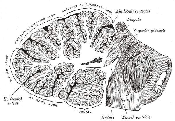
Structure
The cerebellum is composed of two hemispheres, connected by the vermis at the midsagittal plane. It is subdivided into three lobes – anterior, posterior, and flocculonodular, separated by two transverse fissures.
- Anterior lobe – Located above the primary fissure.
- Posterior lobe – Located between the primary fissure and posterior fissure.
- Flocculonodular lobe – Located below the posterior fissure.
Connections
- Superior cerebellar peduncles – midbrain
- Middle cerebellar peduncles – pons
- Inferior cerebellar peduncles – medulla
Zones
Relations
- Superiorly
- Tentorium cerebelli – an extension of the dura mater separating the cerebellum from the cerebrum
- Straight sinus
- Occipital lobe
- Quadrigemnial cistern – contains the great cerebral and pineal gland
- Oculomotor nerve – emerges from brainstem between posterior cerebral artery (above) and superior cerebellar artery (below).
- Anteriorly
- Fourth ventricle
- Pons
- Medulla oblongata
- Superior and inferior medullary velum
- Cerebellopontinge angle cistern
- Inferiorly
- Cisterna magna
- Foramen magnum
- Posterior inferior cerebellar artery

Blood supply
Receives vascular supply from three main arteries originating from the vertebrobasilar system (superior to inferior):
- Superior cerebellar artery (branch of the basilar artery)
- Supplies superior surface and vermis to horizontal fissure
- Gives off superior vermian branch and lateral (marginal) branch
- Anterior inferior cerebellar artery (branch of the basilar artery)
- Supplies anterior inferior surface and middle cerebellar peduncle
- Posterior inferior cerebellar artery (branch of the vertebral artery)
- Supplies posterior inferior surface of cerebellar hemispheres up to horizontal fissure, inferior cerebellar peduncle and inferior vermis
- Gives off inferior vermian branch, choroidal branch to 4th ventricle and cerebellar tonsillar branch
Venous drainage
- Superior (Galenic) group
- drains the midbrain, upper pons, superior vermis and the superior surface of the cerebellar hemispheres
- drains into the vein of Galen and has three named veins
- precentral cerebellar vein
- superior vermian vein
- anterior pontomesencephalic vein
- Anterior (petrosal) group
- drains the lateral pons, medulla and the anterior surface of the cerebellar hemispheres
- drains into the inferior petrosal sinus and has a petrosal vein
- Posterior (tentorial) group
- drains the inferior vermis, tonsils and the posteroinferior surface of the cerebellar hemispheres
- drains into the transverse sinus and has an inferior vermian vein
Variants
- Agenesis
- Tonsillar ectopia – asymptomatic protrusion of tonsils through formen magnum