

Overview
The spleen is a highly vascular, fist-sized solid lymphoid organ (the largest in the body), located in the left hypochondriac intraperitoneal region between the fundus of the stomach and the diaphragm and spanning the 9th to 11th ribs. It functions mainly as a blood filter, sequestering mature red blood cells (red pulp) and providing cell-mediated and humoral immune responses (white pulp)
Structure
- Encased in a weak outer connective tissue capsule which allows for protection and also expansion of the organ
- Surrounded by visceral peritoneum except at the hilum on its visceral surface
- Subdivided into internal sections called lobules
- The spleen has an anterior and posterior segment and rests on the upper pole of the left kidney and tail of the pancreas.
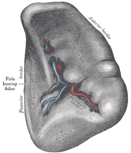
- Hilum – point of entry for splenic vessels, occasionally tail of pancreas reaches this area. Found on the inferomedial aspect of the gastric imprint. The splenic hilum contains nerves, splenic vessels, and also contains attachments for the splenorenal and gastrosplenic ligaments.
Borders
The spleen has has anterior, superior, posteromedial and inferior borders
- The posteromedial and inferior borders are smooth
- The anterior and superior borders contain notches
Surfaces
The spleen has 2 surfaces, the visceral and diaphragmatic.
- Diaphragmatic surface – smooth and convex, in contact with the diaphragm and ribcage
- Visceral surface – concave and irregular with several impressions from contact with abdominal viscera
Impressions
- Gastric imprint – the most concave imprint from the fundus of the stomach.
- Renal imprint – the left kidney leaves an imprint on the intermediate and inferior borders
- Colic imprint – from the splenic flexure of the colon.
- Pancreatic imprint – the tail of the pancreas leaves an impression between the hilum and colic impression sites.
Ligaments
The spleen is connected to the stomach and kidney by parts of the greater omentum (a double fold of peritoneum that originates from the stomach):
- Gastrosplenic ligament – connects hilum of spleen to greater curvature of stomach, contains short gastric and left gastro-omental vessels
- Splenorenal ligament – connects hilum of spleen to left kidney, contains splenic artery and vein
- Between the gastrosplenic ligament and splenorenal ligaments is the lesser sac
- Phrenicocolic ligament – Splenic flexure to diaphragm, supports the spleen inferiorly
- May also be attached to splenic flexure by splenocolic ligament
Arterial Supply
- Splenic artery – major branch of coeliac trunk, travels laterally towards the left and retroperitoneally within the splenorenal ligament, to course behind the pancreas to reach hilum
- The splenic artery branches into five vessels – each supplying different vascular segments of the spleen
Venous Drainage
- Splenic vein – leaves hilum, passes to the right, posterior to the pancreas, to join superior mesenteric vein to become portal vein behind neck of pancreas
Lymphatic Drainage
Follow the splenic artery and vein, and drain into the pancreaticosplenic lymph nodes, and ultimately the coeliac nodes.
Innervation
- Coeliac plexus – from right vagus nerve
Relations
- Diaphragmatic surface faces posterolaterally, related to ribs 9-11 and inferior surface of diaphragm
- Visceral surface faces anteromedially, related to body of stomach, tail of pancreas, upper pole of left kidney, splenic flexure of colon
Development
- Formed in the dorsal mesogastrium
Anatomical Variants
- Splenuculus – accessory spleen
- Wandering spleen (migration from usual position, usually lower pelvis, result of abnormal suspensory ligaments)
- Polysplenia/asplenia
- Retrorenal spleen
- Splenogonadal fusion
Radiological Appearance
CT
- Non-contrast: Homogenous density of 40-60 HU. Hypodense relative to liver.
- C+ Arterial: Zebra spleen (also referred to as psychedelic spleen, tigroid splenic enhancement or more correctly inhomogeneous splenic enhancement), refers to the transient heterogeneous parenchymal arterial enhancement due to the red and white pulp.
- Archiform: alternating bands of low and high density which may look like rings or zebra stripes
- Focal: single area of low density
- Diffuse: mottled appearance
- C+ Portal Venous/Delayed: Homogenous enhancement.
MRI
- T1: Hypointense relative to liver
- T2: Hyperintense relative liver
- DWI: Diffusion restriction.