

Overview
The portal vein supplies the liver with nutrient-rich blood (also containing toxins extracted from digested contents) drained from the gastrointestinal tract, pancreas, spleen and gallbladder. It accounts for 75% of the total blood supply to the liver, with the remainder derived from the hepatic artery proper. The liver is drained by the hepatic veins.
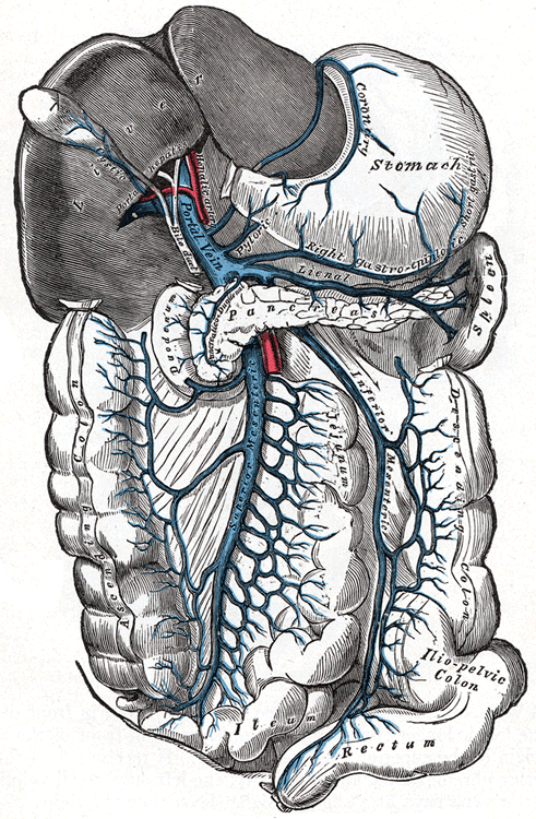
[foogallery id=”5688″]
Origin
The portal vein is formed by the confluence of the superior mesenteric vein and splenic vein, posterior to the neck of the pancreas. The point of origin defines the location of the pancreatic neck and corresponds to the vertebral level of L2.
Course
- From the origin, it courses superiorly towards the right within the hepatoduodenal ligament posterior to the other structures of the portal triad (common hepatic duct and common hepatic artery)
- Anterior to the inferior vena cava, it forms the anterior border of the epiploic foramen (of Winslow).
- At the porta hepatis, the portal vein divides into the left and right portal veins
Tributaries
The splenic vein receives tributaries from the:
- Inferior mesenteric vein – drains the splenic flexure, descending colon, sigmoid colon and superior rectum
- Short gastric veins – drains the fundus of the stomach
- Left gastro-omental vein – drains the greater curvature of the stomach
- Pancreatic veins – drain the pancreas
The portal vein receives tributaries from the:
- Para-umbilical veins – drains the skin of the umbilical region
- Left and right gastric vein – drains the stomach
- Cystic vein – drains the gallbladder
Branches
The typical branching pattern is found in 65% of individuals.
Left Portal Vein
The left portal vein divides into transverse and umbilical portions, delineated by the ligamentum venosum:
- Transverse portions
- Umbilical portions – The main branches of the left portal vein originate from the umbilical portion, and supplies the left hepatic lobe (segments 2, 3 and 4a/4b), the caudate lobe and quadrate lobe.
Right Portal Vein
The right portal vein supplies the right lobe of the liver and divides into:
- Anterior branch – Supplies segments 5 and 8 of the liver
- Posterior branch – Supplies segments 6 and 7 of the liver
Once in the liver, the portal veins ramify, forming smaller venous branches and ultimately portal venules. Each portal venule courses alongside a hepatic arteriole and the two vessels form the vascular components of the portal triad. These vessels ultimately empty into the hepatic sinusoids to supply blood to the liver.
Relations
- Anterior – It lies posterior to the the neck of the pancreas and the first part (D1) segment of the duodenum
- Posterior: It is anterior to the inferior vena cava and the right crus of the diaphragm.
- Left: It is to the right of the common bile duct and hepatic artery proper.
- Right: It is to the left of the foramen of Winslow.
Anatomical Variants
- Portal vein trifurcation (most common) – portal vein divides into the left portal vein, right anterior portal vein and right posterior portal vein
- Right posterior portal vein as the first branch of portal vein
- Segments 6 or 7 supplied by a separate branch of the right posterior branch
- Absent right portal vein (rare)
- Portal vein duplication (rare)
- Absent left extrahepatic portal vein (rare) – A single right portal vein originates from the porta hepatis, supplying the right lobe, then following an intrahepatic course with distalmost branches supplying the left liver
- Circumportal pancreas – The pancreatic parenchyma from the uncinate process is fused with the body of the pancreas, encasing the portal vein and/or the superior mesenteric vein
- Preduodenal portal vein – Main portal vein courses anterior to the duodenum and pancreas, frequently associated with other abnormalities and situs inversus.
Imaging Characteristics
Normal portal vein enhances uniformly in the portal venous phase (60–70 s after intravenous contrast administration), measures 11–13 mm in diameter and 7–8 cm in length.
Since this venous system is valveless, pressure modifications caused by respiration can affect its diameter; therefore, measurements on every imaging technique should be made at deep inspiration, when the calibre is at its greatest.