

Overview
The rectum a midline organ is the most distal segment of the large bowel and acts as a reservoir for faeces. It is macroscopically distinct from colon with absence of taenia coli, haustra and omental appendages.
- Omental appendices – peritoneal covered accumulations of fat on external surface
- Taenia coli – segregation of muscle in its walls into three narrow bands (mesenteric, omental and free)
- Haustra – internal sacculations of the colon
Peritoneum covers anterior and lateral surfaces of the upper third, and anterior surface of middle third. The inferior third is not covered by peritoneum. It is surrounded by fat within a layer of mesorectal fascia, circular extension of endopelvic fascia.
Origin
- Continuous above with sigmoid colon at level S3
Course
- Descends, following the sacral flexure – the anteroposterior curvature of the sacrum (anteriorly concave)
- Three lateral curvatures (superior, intermediate and inferior), formed by the transverse folds of the internal rectum wall
- Anorectal flexure – anteroposterior curve with convexity anteriorly (creating a 90 degree angle), formed by the tone of the puborectalis muscle, and contributes significantly to faecal continence.
- Ampulla – Final dilated segment of the rectum dilated which relaxes to accumulate faeces until defecation
Termination
- Continuous with the anal canal at the pelvic diaphragm
Arterial Supply
- Superior rectal artery
- Terminal continuation of inferior mesenteric artery as it crosses the pelvic brim
- It divides, opposite the third sacral vertebra into two branches (left and right), which descend one on either side of the rectum. About 10 or 12 cm from the anus, these branches break up into several small branches
- Supplies superior two-thirds of rectum
- Middle rectal artery, branch of anterior division of internal iliac artery
- Supplies part of inferior third of rectum
- Inferior rectal artery, branch of internal pudendal artery (derived from anterior division of internal iliac artery)
- Supplies part of inferior third of rectum

Venous Drainage
- Superior rectal vein, tributary of inferior mesenteric vein
- Empties into the portal venous system
- Middle rectal vein, tributary of internal iliac vein
- Empties into systemic venous system
- Inferior rectal vein, tributary of pudendal vein
- Empties into systemic venous system
Importantly, site of portocaval anastomosis – anastomoses between portal and systemic veins are located within the wall of the anal canal
Innervation
Sympathetic
- Lumbar splanchnic nerves (L1 – 2)
- Superior and inferior hypogastric plexuses
Parasympathetic
- Pelvic splanchnic nerves (S2-4) and inferior hypogastric
Lymphatic Drainage
- Superiorly via inferior mesenteric nodes
- Inferiorly via internal iliac nodes
Relations
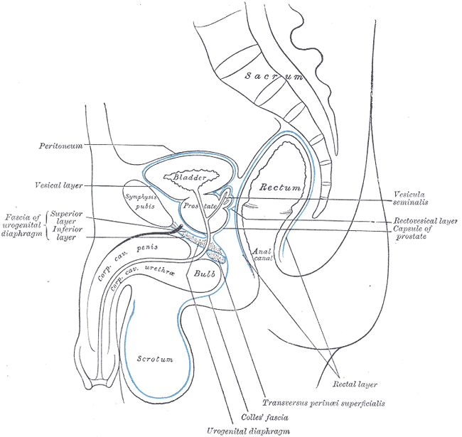
Anterior (male)
- Prostate (separated by Denonvillier’s fascia), seminal vesicles, rectovesical pouch
- Bladder, sigmoid colon, ileum
Anterior (female)
- Vagina (separated by rectovaginal fascia), cervix, rectouterine pouch (of Douglas)
- Bladder, sigmoid colon, ileum

Posterior
- Sacrum, coccyx
- Piriformis, coccygues, levator ani
- Sacral plexus
Superior
- Sigmoid colon
Anatomical Variants
- Middle rectal artery may be absent or arise from internal pundenal or inferior gluteal