

Overview
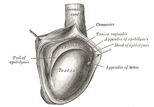
The testes are paired ellipsoid-shaped male sex glands located within the scrotum which have endocrine and exocrine functions. They are suspended from the abdomen by the spermatic cord. The epididymis is situated on the posterolateral aspect of each testicle. The testes play a role in spermatogenesis, while the epididymi play a role in sperm storage and maturation.
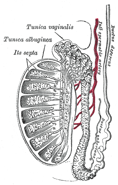
Structure
Testis
- Covered by tunica vaginalis – closed sac of parietal peritoneal origin containing a small amount of viscous fluid
- Testicular parenchyma protected by tunica albuginea which encloses the testes. It penetrates the parenchyma with diaphragms which forms lobules
- Each lobue contains seminiferous tubules supported by interstitial tissue.
- Seminiferous tubules – lined by Sertoli cells responsible for production of spermatozoa
- Interstitial tissue – contain the Leydig cells responsible for testosterone production
- Rete testes – collecting system for spermatozoa
- Efferent tubulents – transport the spermatozoa to the epididymysis for storage and maturation
Epididymis
The epididymis consists of a single heavily convoluted duct, which can be divided into three parts:
- Head – Most proximal part of the epididymis, formed by the efferent tubules of the testes, which transport sperm from the testes to the epididymis.
- Body – Formed by the heavily coiled duct of the epididymis.
- Tail – The most distal part of the epididymis. It marks the origin of the vas deferens, which transports sperm to the prostatic portion of the urethra for ejaculation.
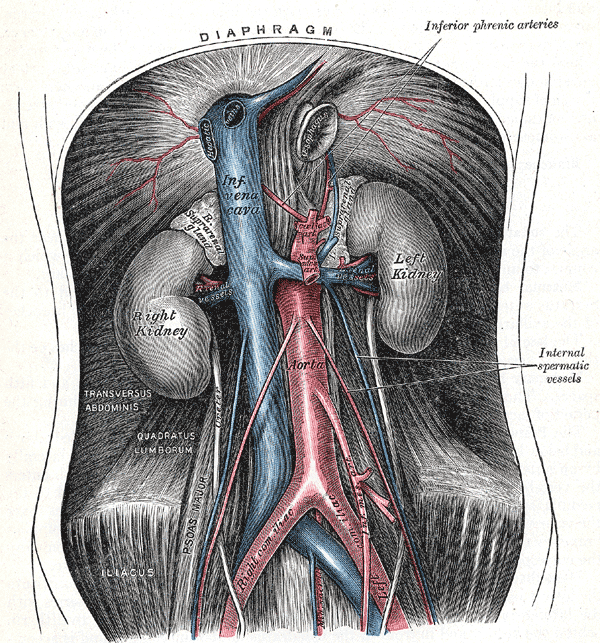
Arterial Supply
- Testicular artery – paired arteries arising directly from the anterolateral aspect of the abdominal aorta at the L2-L3 vertebral levels, distal to the origin of the renal arteries. They descend down the abdomen in the retroperitoneum and cross over the ureter, pass through the deep inguinal ring to enter the scrotum via the spermatic cord. Main supply for both testes and epididymis. It anastomoses with:
- Branches of cremasteric artery – derived from the inferior epigastric artery (arises just before external iliac artery passes under inguinal ligament)
- Branches of artery of the vas deferens – derived from inferior vesical artery (arises from anterior division of internal iliac artery)
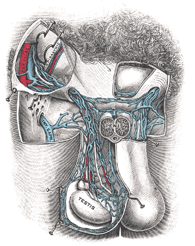
Venous Drainage
Pampniform plexus – formed by the paired testicular veins. The pampiniform plexus wraps around the testicular artery and is anterior to the vas deferens. The veins converge superiorly to form the testicular vein, which ascends in the retroperitoneal space of the abdomen.
Testicular veins
- Left testicular vein drains into the left renal vein
- Right testicular vein drains directly into the inferior vena cava
Lymphatics
Since the testes are originally retroperitoneal organs, the lymphatic drainage is to the lumbar and para-aortic nodes, along the lumbar vertebrae.
- Note: The scrotum drains into the nearby superficial inguinal nodes.
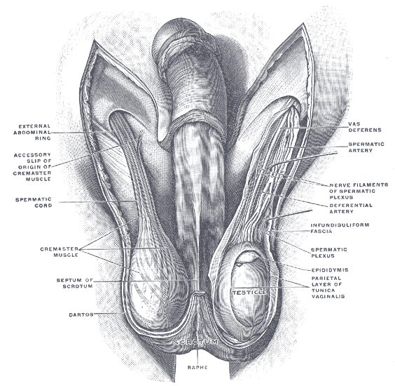
Innervation
- Sympathetic – Lesser splanchnic nerve, synapsing at the coelic ganglion (T10). The post-ganglionic fibres follow the testicular artery to the point of innervation.
- Sensory – genital branch (L2) of the genitofemoral nerve (L1, L2) of the lumbar plexus provides sensory innervation to the tunica vaginalis of the testes.
Anatomical Variants
-
Appendix testis – pair-shaped vestigial structure located at the cranial end of the paramesonephric duct (Mullerian duct), typical at the superior pole in the groove between the head of the epididymis and testis (2%)
- Appendix of epididymis – located at the cranial end of the mesonephric duct one, often projecting from the head of the epididymis. (25%)
- Anterior epididymis – located anterior surface of the testis (7%)