

Overview
The scrotum is a fibromuscular cutaenous dual-chambered sac, located between the penis and anus. The scrotum receives neurovascular supply from the nearby vessels and nerves, in contrast to the testes, which carry their vessels, nerves and lymph drainage from the abdomen during their development.
Structure & Layers
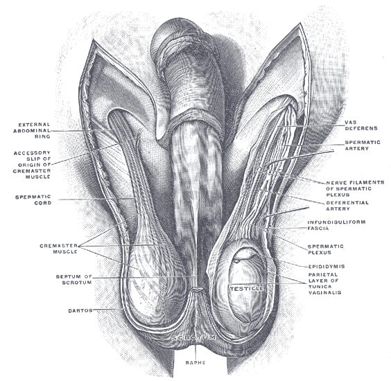
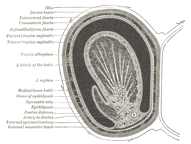
A thin external sac (8 mm), divided into two compartments by the scrotal septum. The scrotal wall and underlying layers are composed of the following structures from superficial to deep:
- Pigmented skin with rugal folds
- Superficial fascia and dartos muscle – a sheet of smooth muscle which helps thermoregulation of the testes by wrinkling the skin, thereby decreasing surface area, reducing heat loss.
- External spermatic fascia – continuation of external oblique abdominis muscle aponeurosis
- Cremasteric fascia – continuation of internal oblique abdominis muscle aponeurosis
- Internal spermatic fascia – continuation of transversus abdominis muscle aponeurosis (transversalis fascia)
- Tunica vaginalis
- Tunica albuginea which covers the testes
Contents
The scrotum contains three major paired structures:
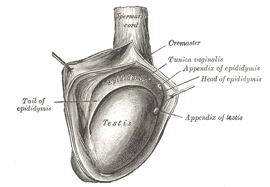
- Testis – paired ellipsoid-shaped male sex glands which play a role in spermatogenesis
- Epididymis – situated at the head of each testicle. Play a role in sperm storage and maturation.
- Spermatic cord – a collection of muscle fibres, vessels, nerves and ducts that run to and from the testes
Arterial Supply
- Anterior scrotal artery – branch of the superficial external pudendal artery, which arises from the medial side of the femoral artery (supplies the labia majora in females)
- Posterior scrotal artery – branch of internal pudendal artery, which arises from the anterior division of internal iliac artery
Venous Drainage
The scrotal veins follow the major arteries, draining into the external pudendal veins.
Innervation
Cutaneous innervation to the scrotum is supplied according to the topography:
- Anterior and anterolateral aspect – The cremaster muscle and anterior scrotum receive innervation from the genital branch of genitofemoral nerve (L1 – L2) and ilioinguinal nerve (L1) (from the lumbar plexus)
- Posterior aspect – Posterior scrotal nerves derived from the perineal branches of the pudendal nerve and posterior femoral cutaneous nerve.
- Cremasteric Reflex: Both the ilioinguinal and the genitofemoral nerves provide a sensory synapse that activates the motor neurons responsible for the cremasteric reflex. This physiologic reflex has both protective and thermoregulatory testicular functions.
Muscles
The muscle fibres contained in the scrotum are the dartos muscle and the cremasteric muscle:
- Dartos Muscle – a smooth muscle sheet located underneath the skin of the scrotum
- Cremasteric Muscle – cremasteric muscle is a paired muscle composed of two parts, a medial cremaster portion originating from the pubic tubercle and the lateral cremaster portion originating from the internal oblique muscle
Lymphatic Drainage
- The lymphatic fluid from the scrotum drains to the nearby superficial inguinal nodes.
- The arrangement of the lymphatic drainage of the scrotum is related to the development of this sac.
- The wall of the scrotum drains into the superficial inguinal lymph nodes.
- The contents of the scrotum drain to the lumbar lymph nodes (as the testes migrate from the abdominal wall through the inguinal canal and into the scrotum, dragging its neurovascular supply and lymph vessels along with it.
Anatomical Variants
- Accessory Scrotum (rare) – most commonly seen mid perineum and does not contain testes
- Bifid Scrotum – an abnormal midline cleft caused by an incomplete fusion of the labioscrotal folds, associated with hypospadias
- Ectopic Scrotum – due to defect in the formation of the gubernaculum. Common sites include suprainguinal, inguinal, infra-inguinal, or perineal regions
- Penoscrotal Transposition (rare) – a genital malpositioning in which the penis is lying in an abnormal location in relation to the scrotum, due to a defect of the scrotum during its caudal migration in the 9th to 11th week of gestation. This inversion can be classified as either partial or complete.
- A partial transposition occurs when the penis lies in the middle of the scrotum.
- A complete penoscrotal transposition is seen when the penis emerges beneath the scrotum. Can present in conjunction with central nervous system, cardiovascular and gastrointestinal tract congenital malformations
https://www.ncbi.nlm.nih.gov/books/NBK549893/