

Overview
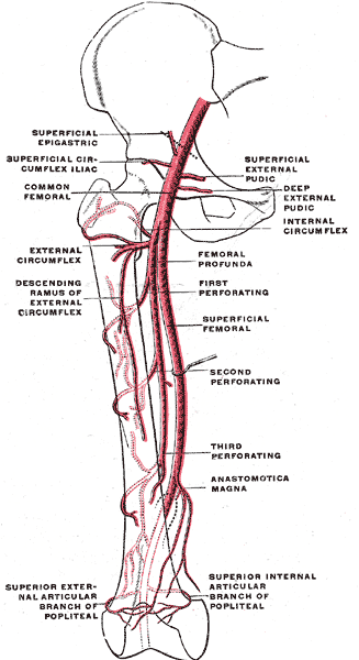
The femoral artery is a continuation of the external iliac artery and is the main arterial supply to the lower extremity. Additionally, it contributes to the blood supply of the anterior abdominal wall. It forms part of a single main trunk that runs from the inguinal ligament to the lower border of the popliteus, where it divides into two branches, the anterior tibial artery and posterior tibial artery. The upper part of the main trunk is named the femoral artery, the lower part the popliteal artery.
Origin
At the midinguinal point (midway between the anterior superior iliac spine and the pubic symphysis), the external iliac artery passes deep to the inguinal ligament into the femoral triangle and becomes the femoral artery.
Segments
- Common femoral artery – The common femoral artery is the segment of the femoral artery between the inferior margin of the inguinal ligament and the branching point of the deep femoral artery
- Superficial femoral (subsartorial) artery – The segment between the branching point of the deep femoral artery and the adductor hiatus, passing through the subsartorial canal.
Course
- In the upper third of the thigh the femoral artery is contained in the femoral triangle (Scarpa’s triangle)
- The first 4 cm of the femoral artery is enclosed with the femoral vein in a funnel-shaped fibrous sheath called the femoral sheath (cural sheath)
- The artery then travels down the anteromedial aspect of the thigh in the femoral triangle giving off branches which supply the anterior thigh muscles
Termination
- Terminates as the popliteal artery as the femoral artery enters the adductor canal (Hunter’s canal) through the adductor hiatus in the adductor magnus, near the junction of the middle and distal thirds of the thigh.
Branches
- Superficial epigastric artery
- Origin: A small branch that arises from the anterior femoral artery, 1 cm distal to the inguinal ligament.
- Course: It passes through the femoral sheath and fascia cribrosa between ascending to cross the inguinal ligament. It ascends between the two layers of the superficial fascia (Camper’s and Scarpa’s) of the anterior abdominal wall towards the region of the umbilicus.
- Supplies: It distributes branches to the superficial subinguinal lymph glands, the superficial fascia and the skin. It anastomoses with branches of the inferior epigastric (branch of the internal thoracic artery) and also with the contralateral superficial epigastric artery
- Superficial circumflex iliac artery
- Origin: A small branch arising just distal to the superficial epigastric artery.
- Course: It pierces the fascia lata and courses laterally parallel with the inguinal ligament towards the region of the anterior superior iliac spine.
- Supplies: It divides into branches which supply the skin overyling the groin, superficial fascia, superficial subinguinal lymph nodes. It anastomoses with the deep iliac circumflex, superior gluteal and lateral femoral circumflex arteries.
- Superficial external pudendal artery
- Origin: A small branch arising from the medial side of the femoral artery.
- Course: It pierces the femoral sheath and fascia cribrosa, coursing medially across the spermatic cord (males)/round ligament (females) and supplies the skin on the lower abdomen, penis, scrotum or labium majus. It anastomoses with the internal pudendal artery (branch of the anterior division of the internal iliac artery).
- Deep external pudendal artery
- Origin: A deeper brancher which courses medially across the pectineus and adductor longus muscle, covered by the fascia lata and piercing the medial side of the thigh.
- Supplies: Supplies the skin of the scrotum or labium majus, perineum and anastomoses with scrotal or labial branches of the perineal artery.
- Deep femoral (profunda femoris) artery
- Origin: A large branch arising from the posterolateral aspect of the femoral artery 2 – 5 cm inferior to the inguinal ligament.
- Course: At its origin, it gives off the medial and lateral femoral circumflex arteries, and during its course it gives off three perforating arteries (first perforating, second perforating and third perforating arteries) which perforate the tendon of the adductor magnus to reach the back of the thigh. It terminates by becoming the fourth perforating artery.
- Medial Femoral Circumflex Artery – arises from the posteromedial aspect of the deep femoral artery, and winds around the medial side of the femur, passing first between the pectineus and psoas major, and then between the obturator externus and the adductor brevis. At the upper border of the adductor brevis it gives off two branches:
- The ascending branch
- The descending branch descends beneath the adductor brevis, to supply it and the adductor magnus; the continuation of the vessel passes backward and divides into superficial, deep, and acetabular branches.
- Lateral Femoral Circumflex Artery – arises from the lateral side of the deep femoral artery, passes horizontally between the anterior and posterior divisions of the femoral nerve, and deep to the sartorius and rectus femoris. It divides into ascending, transverse, and descending branches.
- The ascending branch passes upward, beneath the tensor fascia lata, to the lateral aspect of the hip, and anastomoses with the terminal branches of the superior gluteal and deep iliac circumflex arteries.
- The descending branch runs downward, behind the rectus femoris, upon the vastus lateralis, to which it gives offsets; one long branch descends in the muscle as far as the knee, and anastomoses with the superior lateral genicular branch of the popliteal artery. It is accompanied by the branch of the femoral nerve to the Vastus lateralis.
- The transverse branch, the smallest, passes laterally over the vastus intermedius, pierces the vastus lateralis, and winds around the femur, just below the greater trochanter, anastomosing on the back of the thigh with the medial femoral circumflex, inferior gluteal, and first perforating arteries.
- Descending genicular artery – a small branch that arises from the femoral artery near its termination within the adductor canal. It assists in supplying the knee joint.
- Muscular branches (rami musculares) – arises just before the femoral artery passes through the opening in the tendon of the adductor magnus and divides into a saphenous and musculo-articular branch. The muscular branches supply the sartorius, vastus medialis, and adductores.
- Saphenous branch – anastomoses with the medial inferior genicular artery
- Musculo-articular branch – anatomoses with the medial superior genicular artery and anterior recurrent tibial artery. A branch from this vessel also forms an anastomotic arch with the lateral superior genicular artery.

Relations
- Anteriorly – In the upper part of its course, it is superficial and is covered by skin, superficial fascia, subinguinal lymph glands, superficial iliac circumflex vein, superficial layer of the fascia latera and anterior part of the femoral sheath. In the lower part of its course, it passes behind the sartorius muscle.
- Posteriorly – The artery lies on the psoas, which separates it from the capsule of the hip joint, the pectineus, and the adductor longus. Femoral vein in the lower part of its course intervenes between the artery and the adductor longus.
- Medially – Femoral vein in the upper part of its course.
- Laterally – Femoral nerve and its branches.
Anatomical Variants
- Medial and lateral femoral circumflex arising from the common femoral artery
- Presence of an oblique lateral femoral circumflex artery
- Variable origin of the deep femoral artery (posterolateral most prevalent)