

Overview
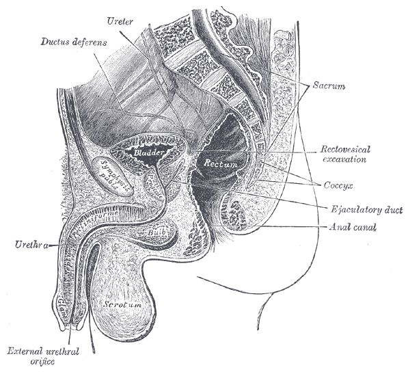
The penis is an external organ of the male reproductive system, located within the urogenital triangle between the perineal membrane superior and the deep perineal fascia inferiorly. It’s primary purpose is for:
- Sexual intercourse
- Micturition – Contains the urethra which transports urine from the bladder to the external urethral orifice
Structure
The penis can be anatomically divided into three parts:
Root
- Most proximal (not externally visible), fixed part of the penis located in the superficial perineal pouch of the pelvic floor.
- The root contains three erectile tissues (two crura and bulb of the penis), and two muscles (ischiocavernosus and bulbospongiosus):
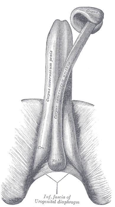
- Left and right crura – located lateral to the bulb of the penis, attached to the ipsilateral ischial ramus and are covered the paired ischiocavernosal muscle
- Bulb of the penis – located in the midline of the penile root, and is traversed by the urethra
- Bulbospongiosus (paired) – associated with the bulb of the penis. It contracts to empty the spongy urethra of any residual semen and urine. The anterior fibres also aid in maintaining erection by increasing the pressure in the bulb of the penis.
- Ischiocavernosus (paired) – surrounds the left and right crura of the penis. It contracts to force blood from the cavernous spaces in the crura into the corpora cavernosa – this helps maintain erection.
Body
- Located between the root and glans, suspended from the pubic symphysis.
- Lacks muscle and consists mostly of three cylinders of erectile tissue – two corpora cavernosa (dorsally) and corpus spongiosum (ventrally).
- It contains the spongy urethra with support skin, connective tissue, blood, lymph and fascia.
- Corpora cavernosa (paired, dorsal) – the anterior continuation of the left and right crura into the dorsal part of the penis. The two erectile tissue masses are separed by the septum of the penis (although incompletely).
- Corpus spongiosum (ventral) – the anterior continuation of the the bulb. The spongy urethra runs through the corpus spongiosum. The corpus spongiosum fills to a reduced pressure during erection to prevent it becoming occluded.
Glans
- Distal part of the of penis, conical in shape and formed by the distal expansion of the corpus spongiosum. The proximal portion of the glans is termed the corona, where the neck of the corona glans separates it from the body of the penis. The tip of the penis contains the urethral meatus (external urethral orifice).
Fascial Coverings
Each mass of erectile tissue has two fascial coverings.
- Colles fascia (external/most superficial fascia) – located immediately under the skin, which is in continuity with the fascia of Scarpa which covers the abdominal wall.
- Buck’s fascia (deep fascia of the penis) – a continuation of the deep perineal fascia. Forms a strong membranous covering which holds all three erectile tissues together.
- Tunica albuginea – (deepest, lies underneath the deep fascia). An individual capsule around each cavernous body and fused in the midline. The incomplete septum between the two corpora is comprised of tunica albuginea.
Skin & Ligaments
The skin of the penis is more heavily pigmented than that of the rest of the body. It is connected to the underlying fascias by loose connective tissue. The root of the penis is supported by two ligaments, which attach it to the surrounding structures:
Ligaments
- Suspensory ligament – a condensation of deep fascia. It connects the erectile bodies of the penis to the pubic symphysis.
- Fundiform ligament – a condensation of abdominal subcutaneous tissue. It runs down from the linea alba, surrounding the penis like a sling, and attaching to the pubic symphysis.
Prepuce (foreskin)
- Double layer of skin and fascia, located at the neck of the glans and covers the glans to a variable extent.
- Connected to the surface of the glans by the frenulum, a median fold of skin on the ventral surface of the penis. The potential space between the glans and prepuce is termed the preputial sac.
Arterial Supply
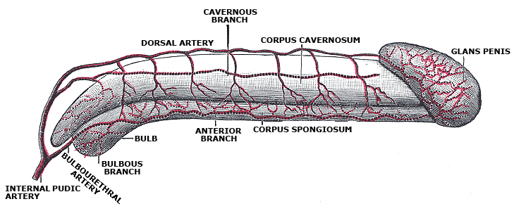
The penis receives arterial supply from three sources which are all branches of the internal pudendal artery (arising from the anterior division of the internal iliac artery)
- Dorsal arteries of the penis
- Deep arteries of the penis
- Bulbourethral artery
Venous Drainage
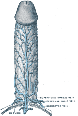
Venous blood is drained from the penis by paired veins.
- Deep dorsal vein of the penis – drains the cavernous spaces and empties into the prostatic venous plexus (Santorini’s) which later drains into the internal iliac vein.
- Superficial dorsal veins – drains the superficial structures of the penis, such as the skin and cutaneous tissues
Lymphatic Drainage
- Anterior urethra drains into deep and superficial inguinal nodes
- Posterior urethra drains into obturator and internal iliac nodes
Innervation
- Erection occurs via parasympathetic innervation which allows engorgement of the corporal bodies – cavernous nerves from prostatic nerve plexus
- Ejaculation occurs via sympathetic response and occurs when semen exits the vas deferens to the ejaculatory ducts where fluid from the seminal glands are added – pudendal nerve
- The penis is supplied by S2-S4 spinal cord segments and spinal ganglia.
- Sensory and sympathetic innervation to the skin and glans penis is supplied by the dorsal nerve of the penis, a branch of the pudendal nerve (sacral plexus).
Anatomical Variants
- Epispadias – defect of the dorsal wall of the urethra
- Hypospadias – urethra opens on the underside of penis
- Anterior and posterior urethral valves
- Duplication
- Congenital stricture