

Overview
Arterial supply to the foot can be divided into plantar and dorsal components, derived from the terminal branches of posterior tibial artery and anterior tibial artery.

Origin
Terminal continuation of the popliteal artery, between the tibia and fibula at the level of the lower margin of the popliteus muscle
Course
- Descends in an inferomedial course through the posterior (flexor) compartment of the leg, accompanied by two veins and by the tibial nerve, which initially lies medially but soon crosses it posteriorly (remaining laterally for the the majority of its course)
- In the lower third of its course, it lies more superficially
- Enters the foot passing inferiorly to the medial malleolus, through the tarsal canal.
- Midway between the malleolus and tubercle of the calcaneus, it terminates by bifurcating into the lateral and medial plantar arteries
Branches
Fibular (Peroneal) Artery
- Deeply seated on the back of the fibular side of the leg
- Arises from the posterior tibial 2.5 cm below the inferior border of the popliteus
- It courses laterally, in an oblique orientation before descending along the medial side of the fibula, contained in a fibrous canal between the tibialis posterior and flexor hallucis longus
- It then runs behind tibiofibular syndesmosis and divides into the lateral calcaneal branches
- It is covered in the upper part of its course by the soleus and deep transverse fascia of the leg, below by the flexor hallucis longus
Termination
Terminates inferior to the medial malleolus as the medial and plantar arteries.
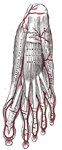
Medial Plantar Artery
- Smaller than the lateral plantary artery
- Courses forward along the medial side of the foot
- At the base of the first metatarsal it passes along the medial border of the first toe, anastomosing with the first dorsal metatarsal artery.
- Small superficial digital branches accompany the digital branches of the medial plantar nerve and join the plantar metatarsal arteries of the first three spaces.
Lateral Plantar Artery
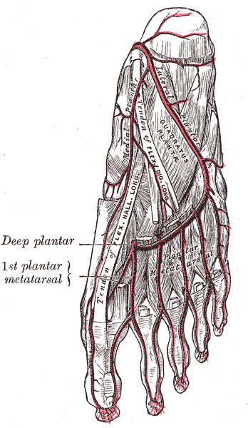
- Larger than the medial plantar artery
- From the medial side of the foot, it the courses laterally in an oblique orientation towards the base of the fifth metatarsal bone
- It then curves medially to the interval between the bases of the first and second metatarsal bones, where it unites with the deep plantar branch of the dorsalis pedis artery, thus completing the plantar arch
Relations
Medial
- Within the tarsal tunnel – tendon of tibialis posterior, tendon of flexor digitorum longus, tibial vein
- Covered by the lacinate ligament
Lateral
- Within the tarsal tunnel – tibial nerve, tendon of flexor hallucis longus
Deep
- Tibialis posterior, flexor digitorum long, tibia, back of ankle joint
Superficial
- Deep transverse fascia of the leg
- Gastrocnemius (most superficial), soleus
Anatomical Variants
- Hypoplastic infrapopliteal vessels (i.e. posterior tibial artery and anterior tibial artery)
- Trifurcation (no tibioperoneal trunk)
- High bifurcation (above popliteus, normally bifurcates at lower border)
- High origin of anterior tibial artery
- High origin of posterior tibial artery
- Fibular artery arising off the anterior tibial artery
- Anterior tibiofibular trunk