

Overview
Arterial supply to the foot is derived from three primary sources: peroneal (fibular), posterior tibial and anterior tibial arteries.

Dorsal Foot
Arterial supply of the dorsal foot arises from the anterior tibial artery. As the anterior tibial artery crosses beneath the superior extensor retinaculum, then the Y-shaped inferior extensor retinaculum of the ankle joint, it becomes known as the dorsalis pedis artery. The dorsal and plantar vasculature are interconnected, mainly via the anastomosis formed by the deep plantar (deep perforating artery) joining the plantar arch – known as the pedal plantar loop. Other anastomosing arterys include the perforating branches arising from the metatarsal arteries which anastomose with the plantar arch and the medial tarsal artery which anastomoses with the medial plantar artery.
Dorsalis pedis artery
- Origin: Continuation of the anterior tibial artery as it passes beneath the Y-shaped inferior extensor retinaculum of the ankle joint.
- Course:
- Several small arteries arise as it moves distally in the direction of the first metatarsal space (anterior lateral malleolar, medial malleolar, lateral tarsal, and medial tarsal arteries)
- It then courses a short distance to the base of the first intermetatarsal space, where it gives off the deep plantar (deep perforating) artery which anastomoses with the plantar arch
- As the dorsalis pedis artery continues further distally, it reaches the tarsal-metatarsal joint line near the middle cuneiform bone and gives off the arcuate artery and the first dorsal metatarsal artery
- Relations: Runs lateral to extensor hallucis longus muscle
First dorsal metatarsal artery
- Continuation of the dorsalis pedis artery
- Supplies dorsal digital branches to the medial and lateral side of the great toe, first cleft and medial side of the 2nd toe
Medial and latareral tarsal arteries
- Branches of the dorsalis pedis artery
- Medial
- Ramifies on the medial border of the foot and joins the medial malleolar arterial network
- Lateral
- Travels laterally, lying beneath the extensor digitorum brevis muscle, supplying it and the underlying tarsal bones
- Anastomoses with perforating branch of the fibular artery (taking part in anastomosis around ankle joint)
Arcuate artery
- The arcuate artery arcs medially-to-laterally over the dorsum of the foot, running beneath the tendons of extensor digitorum brevis muscle, over the bases of the metatarsal bones
- It gives rise to three branches which course within intermetarsal space: second, third, and fourth dorsal metatarsal arteries
- Distally it anastomoses with the lateral tarsal artery
Dorsal metatarsal arteries
- There are three metatarsal arteries which arises as branches of the arcuate artery, and travel in the lateral three clefts, giving off proximal and distal perforating branches that communicate with the plantar metatarsal arteries of the plantar arch
- Second and third dorsal metatarsal arteries – As they travel toward the phalanges, each split into two dorsal digital arteries to create a total of four dorsal digital arteries which supply the second, third, and fourth toes
- Fourth dorsal metatarsal artery – Splits into three dorsal digital arteries in order to supply the fourth and fifth toes.
Plantar foot
The posterior tibial artery supplies the plantar aspect of the foot. As the posterior tibial artery curves around the medial malleolus through the tarsal tunnel, it gives rise to three separate arterial branches (medial calcaneal, medial plantar and lateral plantar artery.
Medial Calcaneal Artery
- Anastomoses with the lateral calcaneal artery (arising from the peroneal (fibular) artery of the leg, forming a vascular network that supplies the medial portion of the calcaneus.
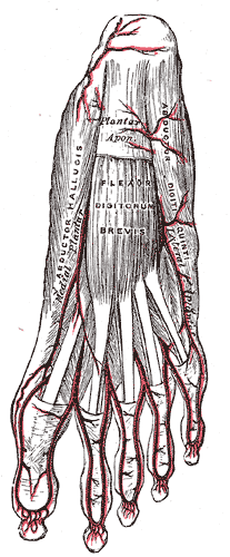
Medial plantar artery
- A smaller calibre vessel supplying the medial side of the foot, abductor hallucis muscle and flexor digitorum brevis muscle
- Course:
- Travels distally through the medial aspect of the foot between the abductor hallucis (superficially) and tendons of the flexor digitorum longus (deep)
- It then splits into deep and superficial branches near the tarsal-metatarsal joint line.
- Branches:
- Deep branch of the medial plantar artery – continues distally toward the hallux, joins with the first plantar metatarsal artery, and supplies the first toe as a plantar digital artery.
- Superficial branch of the medial plantar artery – connects to the circulation of the dorsal foot via the medial tarsal arteries.
- Cutaneous branches – perforate the plantar aponeurosis between abductor hallucis muscle and flexor digitorum brevis muscle
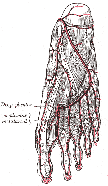
Lateral plantar artery
- Larger calibre vessel
- Course:
- Courses diagonally toward the base of the 5th metatarsal bone between the flexor digitor brevis (superficially) and the quadratus plantae muscle (deep)
- It then splits into two vessels at the level of the proximal fifth metatarsal bone, into superficial branch of the lateral plantar artery and dontinues as a deep trunk to form plantar arch.
- Branches:
- Superficial branch of the lateral plantar artery – travels distally toward the fifth phalanx to become a plantar digital artery that will supply the fifth toe.
- Cutaneous branches – perforate the plantar aponeurosis between flexor digitorum brevis and abductor digiti minimi muscles
Plantar arch
- Course: Arcs medially toward the first metatarsal bone, travelling across the bases of the fourth, third and second metartasal and giving rise to five plantar metatarsal arteries
- Anastomosis of the lateral plantar artery and dorsalis pedis artery
- Lies in the neurovascular plane deep to the plantar aponeurosis, superficial to the long tendons (between the first and second anatomical layers)
- Joins the dorsalis pedis artery in the proximal part of the first intermetatarsal space via the deep perforating artery
Plantar metatarsal arteries
- The are four plantar metatarsal arteries which arise from the plantar arch and run forward between the metatarsal bones, supplying the four clefts and digits
- It anastomoses with the dorsal metatarsal arteries via perforating arteries
- The first four of these plantar metatarsal arteries travel distally through the metatarsal spaces, and each split into two dorsal digital arteries to create a total of eight (which supply all 5 toes)
- Final plantar metatarsal artery (i.e. first plantar metatarsal artery) – joins the medial plantar arter
Anatomical Variants
- Absence of the dorsalis pedis artery: dorsal supply from the plantar arteries
- Dorsalis pedis as the continuation of the peroneal artery
- Hypoplastic dorsalis pedis
- Absence of the arcuate artery
- Arcuate artery arising from the lateral tarsal artery
- Supply of the dorsal metatarsal arteries from the plantar arch alone
- No communication between the plantar and dorsal arch