

The anterolateral abdominal wall encloses the abdominal cavity and consists of four main layers (external to internal): skin, superficial fascia, muscles and associated fascia, and parietal peritoneum. It has multiple functions:
- Forms boundary which keeps the abdominal viscera in the abdominal cavity and assists the viscera in maintaining their anatomical position against gravity.
- Protects the internal abdominal viscera from injury.
- Assists in forceful expiration by pushing the abdominal viscera upwards, thereby increasing intra-abdominal pressure

Boundaries
- Superior: xiphoid process of the sternum and costal margins
- Posterior: vertebral column
- Inferior: pelvic bones and inguinal ligament
Layers
-
Skin
-
Superficial fascia
-
Above umbilicus – a thin single sheet of connective tissue continuous with the superficial fascia in other regions of the body.
- Below the umbilicus – divided into two layers; the fatty superficial layer (Camper’s fascia) and the membranous deep layer (Scarpa’s fascia). The superficial vessels and nerves run between these two layers of fascia.
-
-
Muscle
-
Fascia
-
Parietal peritoneum
Flat Muscles
The muscles of the anterolateral abdominal wall can be divided into two main groups:
- Flat muscles – three flat muscles, situated laterally on either side of the abdomen, with fibres running in differing directions to each other. Each flat muscle forms an aponeurosis (a broad, flat tendon), which covers the vertical rectus abdominis muscle. These aponeuroses entwine in the midline of the anterior abdominal wall, forming the linea alba (a fibrous structure that extends from the xiphoid process of the sternum to the pubic symphysis).
- Vertical muscles – two vertical muscles, situated near the mid-line of the body.
External Oblique
The largest and most superficial flat muscle. Its fibres run inferomedially.
- Origin: Originates from ribs 5-12
- Insertion: Iliac crest and pubic tubercle
- Functions: Contralateral rotation of the torso
- Innervation:
- Thoracoabdominal nerves (T7-T11) – anterior continuation of the intercostal nerves T7-T11 from the intercostal spaces into the abdominal wall. They travel between the internal oblique and transverse abdominis to the sheath of the rectus abdominis which they perforate.
- Subcostal nerve (T12) – rungs along the border of the 12th rib
Internal Oblique
The internal oblique lies deep to the external oblique. It is smaller and thinner in structure. Its fibres run superomedially (perpendicular to the fibres of the external oblique).
- Origin: Originates from the inguinal ligament, iliac crest and lumbodorsal fascia
- Inserts: Ribs 10-12
- Functions: Bilateral contraction compresses the abdomen, while unilateral contraction ipsilaterally rotates the torso.
- Innervation:
- Thoracoabdominal nerves (T7-T11)
- Subcostal nerve (T12)
- Branches of the lumbar plexus
Transversus Abdominis
The deepest of the flat muscles. Deep to this muscle is the transversalis fascia. Its fibres run transversely.
- Origin: Inguinal ligament, costal cartilages 7-12, the iliac crest and thoracolumbar fascia.
- Inserts: Conjoint tendon, xiphoid process, linea alba and the pubic crest.
- Functions: Compression of abdominal contents.
- Innervation: Thoracoabdominal nerves (T7-T11), subcostal nerve (T12) and branches of the lumbar plexus.
Vertical Muscles
There are two vertical muscles located in the midline of the anterolateral abdominal wall – the rectus abdominis and pyramidalis.
Rectus Abdominis
Long, paired muscles found either side of the midline in the abdominal wall, split by the linea alba. The lateral borders of the muscles create a surface marking known as the linea semilunaris. At several places, the muscle is intersected by fibrous strips, known as tendinous intersections.
- Origin: Crest of the pubis
- Inserts: Xiphoid process of the sternum and the costal cartilage of ribs 5-7.
- Functions: Assists the flat muscles in compressing the abdominal viscera. Also stabilises the pelvis during walking, and depresses the ribs.
- Innervation: Thoracoabdominal nerves (T7-T11)
Pyramidalis
A small triangular muscle found superficial to the rectus abdominis near the base of the pubis bone. The apex of the triangle attached to the linea alba.
- Origin: Pubic crest and pubic symphysis
- Inserts: Linea alba
- Functions: It acts to tense the linea alba
- Innervation: Subcostal nerve (T12).
 Surface Anatomy
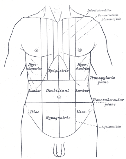
The anterolateral abdominal wall can be divided into several topographical areas, useful for clinically describing location of signs and symptoms:
- Four quadrants system – Divided by the horizontal transumbilical and vertical median planes, resulting in four quadrants termed right upper, left upper, right lower and left lower quadrants.
- Nine abdominopelvic regions system –
- Divided horizontally by the superior subcostal plane (under the costal margins of the 10th ribs) or transpyloric plane (midpoint between jugular notch and pubic symphysis, at level of L1 vertebrae) and the inferior intertubercular plane (connects the tubercules of the iliac crest).
- Divided vertically by the two midclavicular planes which pass through the midpoint of each clavicle and the mid-inguinal point (halfway between the pubic symphysis and the anterior superior iliac spine).
- The four planes form nine abdominal regions termed hypochondriac (right, left) and epigastric regions superiorly, flanks (right, left) and umbilical region in the middle, groin (right, left) and hypogastric region inferiorly.
Arterial Supply
Above Umbilicus
- Superior epigastric arteries
- Origin: terminal branches of internal thoracic arteries at the level of the 6th or 7th costal cartilage
- Course: descends to the anterior abdominal wall, pierces diaphragm to run in rectus sheath anterior to the transversus abdominis, behind the rectus muscle.
- Branches: branches of superior epigastric artery perforate the rectus sheath and supply the skin of the abdomen
- Supplies: anterior abdominal wall, skin of the abdomen
- Anastomosis with inferior epigastric arteries
- Inferior epigastric arteries
- Origin: external iliac artery superior to the inguinal ligament and medial to deep inguinal ring
- Course: bends in an oblique path passing along the medial border of the deep inguinal ring, it then punctures the transveraslis fascia and enters the rectus sheath just beneath the arcuate line
- Termination: anastomoses with the superior epigastric artery above the umbilicus
- Branches: cremasteric artery, pubic branch and muscular branches
- Supply: rectus abdominis muscle; deep abdominal wall of the pubic and lower umbilical regions
- Relations: accompanied by the inferior epigastric vein and is crossed by the ductus deferens or round ligament of the uterus
- Subcostal
- Origin: Arises from the distal descending thoracic aorta
- Course: Travels beneath the 12th rib in the subcostal space to enter the abdomen.
- Pierces transversus abdominus aponeurosis to anastomose with superior epigastric.
Below Umbilicus
- Branches of Common femoral artery
- Superficial epigastric
- Superficial circumflex iliac
- Superficial Inferior Epigastric
- Deep circumflex iliac
Venous Drainage
- Above umbilicus – azygous system
- Below umbilicus – great saphenous vein
Lymphatic Drainage
- Above umbilicus – axillary and parasternal nodes
- Below umbilicus – superficial inguinal nodes
- Deep – follows arterial supply to external iliac, parasternal and posterior mediastinal nodes
Innervation
- Motor and sensory innervation by anterior rami of spinal nerves T7-L1 as intercostal nerves
- 7-11, subcostal, iliohypogastric and ilioinguinal nerves
Anatomical Variants
Inferior epigastric artery
- Can arise beneath the inguinal ligament from the femoral artery or profunda femoris artery
- Can arise from a common trunk with the obturator artery arising off the external iliac artery
- Can be doubled
- Can rarely arise from the obturator artery