Overview
Large, thick bone with an inverted triangular shape located at the terminal vertebral canal, forming the posterior aspect of the pelvis. It transmits the total body weight between the lower appendicular skeleton and the axial skeleton. Formed by the fusion of 5 sacral vertebrae.


Gross Anatomy
- Base – articulates with 5th lumbar vertebra via intervertebral disc
- Apex – articulates with coccyx inferiorly
- Ala – expanded wing-like transverse processes, fused between sacral vertebra creating four pairs of anterior and posterior sacral foramina
- Promontory – prominent anterior edge of body of S1
- Central canal – continues along the core of sacrum, ending in 4th sacral foramina as the sacral hiatus.
- Sacral foramina – transmits anterior and posterior rami of spinal nerves S1 – 4
- Cornua – inferior projections, articulate with cornua of coccyx
Surfaces

Dorsal Surface
Convex, coarse and rugged, giving rise to three bony ridges:
- Median sacral crest
- Central ridge of bone, formed by fusion of spinous process of first three sacral vertebrae.
- Gives attachment to the supraspinous ligament
- Intermediate sacral crest
- Formed by the fusion of sacral articular processes
- Gives attachment to posterior sacroiliac ligaments
- Lateral sacral crest
- Formed by the fusion of transverse processes
- Gives attachment to the sacrotuberous ligament and posterior sacroiliac ligaments
Pelvic Surface
- Marked by four transverse lines – the remnants of the fused sacral intervertebral discs (fusion of the sacral vertebrae begins at age 20).
- Sacral promontory – anterior projection of S1 which forms the posterior margin of the pelvic inlet. It is continuous with the margin of the ala of the sacrum, arcuate line of the ilium, and the pectin pubis and pubic crest of the pubic bone.

Auricular Surface
Bilateral outer-ear shaped surfaces which articulate with the ilium

Muscular Attachments
Anterior Surface
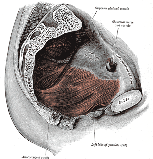
- Piriformis
- Flat pyramidal, lateral rotator muscles
- Originates from S2 – S4 level of the pelvic surface and attaches to the greater trochanter of the femur
- Externally rotates, abducts, extends and stabilises the hip joint
- Coccygeus
- Inserts on the lower sacrum
- Gives support to the contents of the pelvic cavity and due to its attachment to the coccyx, is able to flex the bone.
- Iliacus
- Arises from the iliac fossa but also has fibres originating at the ala of the sacrum.
- Distal attachment to the lesser trochanter of the femur as the iliopsoas
- Flexes the thigh at the hips and stabilise the hip joint.
Posterior Surface
- Gluteal maximus
- Multifidus lumborum
- Deepest muscle
- Attaches to the transverse processes of the superior vertebrae
- Helps stabilise the spine
- Erector spinae
- Partly arises from the posterior sacrum and the sacrospinous ligament.
- Extends and laterally bends the head and vertebral column.
Innervation

Spinal Cord
- Central canal contains sacral fibres of the cauda equina
- The proximal parts of these fibres are contained within the dural sac, which terminates at about the level of S2.
- The filum terminale (a continuation of the pia mater from the conus medullaris of the spinal cord at L2) is joined by the arachnoid and dura mater at the level of S2 and continues inferiorly through the sacrum as the coccygeal ligament to its attachment at the coccyx. It is surrounded by the expanded sub-arachnoid space which forms the lumbar cistern.
Autonomic Nerves
The sacral part of the two sympathetic trunks run along the pelvic surface of the sacrum, medial to the sacral foramina. Each trunk has four ganglia in this region.
Arterial Supply
- Median sacral artery
- Branch of abdominal aorta that arises posteriorly before its bifurcation.
- Runs along midline to the coccyx supplying the posterior rectum, glomus coccygeum (coccygeal gland), and anastomosing with the lateral sacral arteries and supply meninges and sacrum along the way.
- Lateral sacral arteries
- Pair of bilateral vessels that arises from the posterior division of internal iliac artery
- Runs along the medial border of the sacral foramina
- Gives rise to superior and inferior branches
- Supply the meninges, sacrum and surrounding muscles
Relations
Anteriorly:
- Fascia of Waldeyer, peritoneum (S1-S2), rectum (S3-S5), lymph nodes
- in the midline from above down:
- median sacral vessels
- superior rectal artery
- on each side of the midline
- sympathetic chain
- lateral sacral vessels
- piriformis
- sacral plexus
- coccygeus
- levator ani
Variants
- Angel-wing sacrum
- Accessory sacroiliac joints
- Complete agenesis of the dorsal wall of the sacral canal
- Developmental defects in the ala of the sacrum
- Developmental absence of a portion of the right ala of S1
- Midline cleft of S1
- Sacral ribs
- Spina bifida occulta of S1
- Transitional lumbosacral vertebra