

Overview
The hip joint is a ball and socket synovial joint, formed by the articulation of the femoral head with the pelvic acetabulum. This femoroacetabular articulation connects the axial skeleton with the lower extremity for stability and weight-bearing of the upper body.
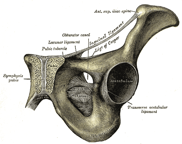
Structures
Acetabulum
- The acetabulum is a cup-like depression located on the inferolateral aspect of the pelvis bilaterally.
- The cavity it forms is deepened by a circumferential fibrocartilaginous collar called the labrum, which confers additional stability by increasing coverage of the femoral articular surface and limits extreme range of motion.
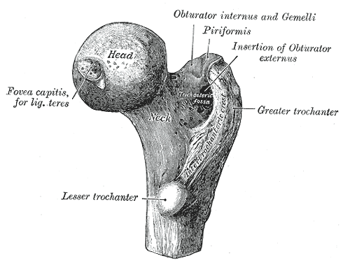
Femoral Head
- The femoral head is a hemispherical articulating surface congruent with the concavity of the acetabulum.
- Both the acetabulum and head of femur is covered by articular cartilage, thicker at points of weight bearing.
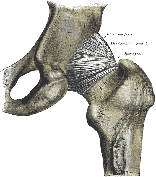
Capsule
- The hip joint capsule is formed by three major ligaments which run in a spiral fashion, preventing hip extension and are surrounded by thick longitudinal fibres that provide additional stability the lateral plane.
- The capsule is thicker anterosuperiorly, where the predominant stresses of weight bearing occurs.
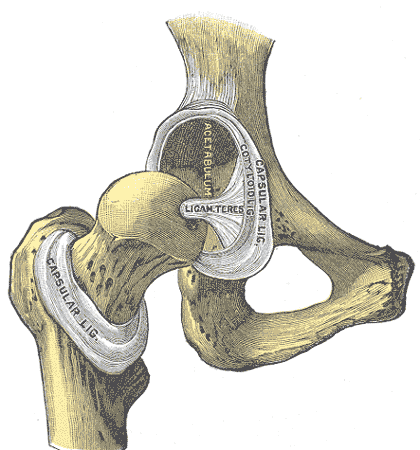
Ligaments
Intracapsular
- Ligamentum teres
- A relatively small ligament which runs from the acetabular fossa to the fovea of the femur
- It has a pyramidal shape with a broad origin
- It encloses a branch of the obturator artery (arises from anterior division of the internal iliac artery)
Extracapsular
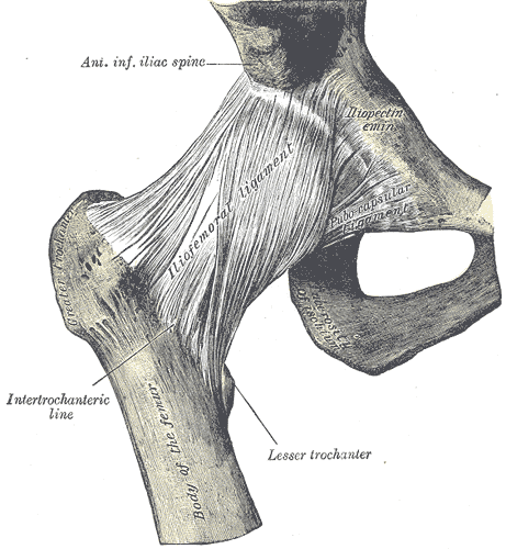
- Iliofemoral ligament (Y ligament of Bigelow)
- Arises from the anterior inferior iliac spine and then bifurcates before inserting into the intertrochanteric line of the femur.
- It has a ‘Y’ shaped appearance, and prevents hyperextension and external rotation of the hip joint.
- It is the strongest of the three ligaments and the strongest ligament in the body
- Pubofemoral
- Spans between the superior pubic rami and the anterior surface of the intertrochanteric line of the femur, often blending with the inferior fibers of the iliofemoral ligament and reinforcing the capsule anteriorly and inferiorly.
- It has a triangular shape, and prevents excessive abduction and extension.
- Ischiofemoral
- It attaches to the posterior surface of the acetabular rim and labrum and courses circumferentially around the joint to its insertion on the anterior aspect of the femur.
- It has a spiral orientation and limits internal rotation and hip adduction with flexion.
Movements & Muscles
No muscles attach directly to the femoral head but rather cross the hip joint.
- Flexion – iliopsoas, rectus femoris, sartorius, pectineus
- Extension – gluteus maximus, semimembranosus, semitendinosus and biceps femoris (the hamstrings)
- Abduction – gluteus medius, gluteus minimus, piriformis and tensor fascia latae
- Adduction – adductors longus, brevis and magnus, pectineus and gracilis
- Lateral rotation – biceps femoris, gluteus maximus, piriformis, assisted by the obturators, gemilli and quadratus femoris.
- Medial rotation – anterior fibres of gluteus medius and minimus, tensor fascia latae
The degree to which flexion at the hip can occur depends on whether the knee is flexed – this relaxes the hamstring muscles, and increases the range of flexion.
Extension at the hip joint is limited by the joint capsule and the iliofemoral ligament. These structures become taut during extension to limit further movement.
Arterial Supply
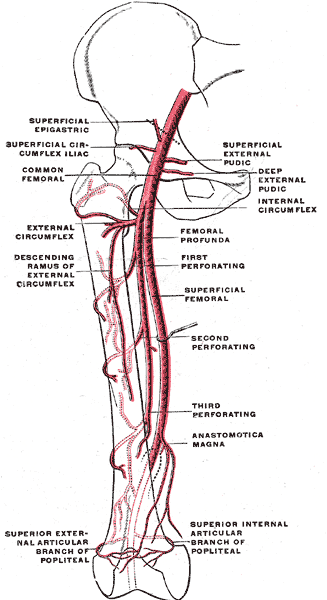
Medial and lateral circumflex femoral arteries
- Branches of the deep femoral artery which anastomoses around the base of the femoral neck to form a ring from which smaller arteries arise to supply the hip joint.
- The medial circumflex femoral artery is responsible for the majority of the arterial supply as the lateral circumflex femoral artery has to penetrate through the thick iliofemoral ligament. Hence, damage to the medial circumflex femoral artery can result in avascular necrosis of the femoral head.
- The lateral epiphyseal branch of the medial femoral circumflex and the ascending branch of the lateral femoral circmflex
- The ligamentum teres artery descends from the posterior branch of the obturator artery and attaches at the fovea
Innervation
Innervated primarily by the sciatic, femoral and obturator nerves. These same nerves innervate the knee, which explains why pain can be referred to the knee from the hip and vice versa.
Development
- The pelvis is made up of three parts: the ilium, ischium, and pubis.
- These innominate bones come together at the triradiate cartilage to form the cup-shaped socket known as the acetabulum.
- At 15 to 17 years of age, the triradiate cartilage begins to ossify, and it completely fuses by 20 to 25 years of age.
- Acetabular development is a complex process that involves both endochondral growth from the triradiate cartilage and intramembranous growth from primary and secondary ossification centers of the innominate bones
Anatomical Variants
- Synovial herniation pits (Pitt pits) – round radiolucencies surrounded by thin sclerotic margins, found predominantly at the superior portion of the proximal anterior femoral neck
- Os acetabuli – formed from the fusion of multiple primary and accessory ossification centre
- Communication between the iliopsoas bursa and the hip joint – the iliopsoas bursa which separates the iliopsoas tendon from the anterior capsule of the hip joint is normally collapsed, however a congenital connection may result in bursal distension by synovialfluid
- Accessory iliacus tendon (~66%)
- Congenital absence of the ligamentum teres
- Hip plicae – synovial folds located at the interface of articular surfaces representing embryonic remnants
- Pectinofoveal fold (middle retinauculum of Weitbrecht) – extends from the lesser trochanter to the fovea capitis and contains the posteroinferior retinacular arteries and branches of the medial femoral circumflex artery
- Stellate crease (or stellate lesion) – a bare area within the acetabular articular surface deficient of hyaline cartilage, located above the anterosuperior margin of the acetabulum and medial to the supraacetabular fossa
- Accessory superior acetabular notch
- Inferior gluteal artery (branch of the anterior division of internal iliac artery) is the main blood supply to the femoral head in a small portion of patients
https://www.ajronline.org/doi/10.2214/AJR.12.9861