

Overview
The femur is the longest, heaviest and strongest bone in the body, and the only long bone in the thigh, serving as attachment site for muscles and ligaments which move the hip and knee joint.
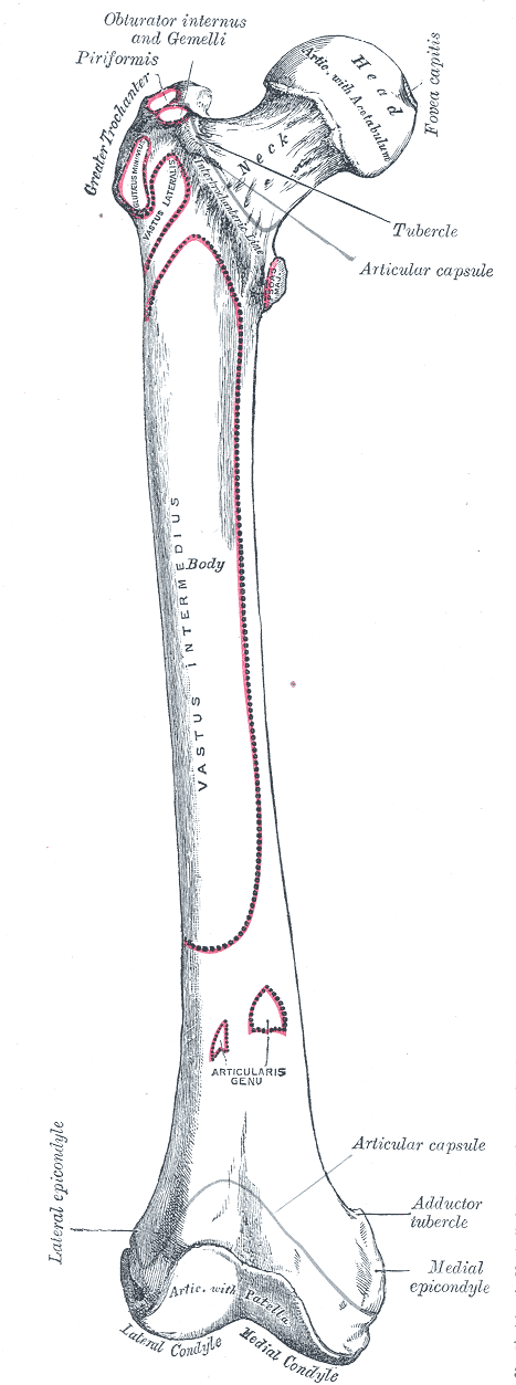
Structure
Head
- Spherical, covered with articular cartilage
- Projects superomedially and articulates with the acetabulum of the pelvis to form the hip (femoroacetabular) joint
- Fovea, small depression – for attachment of the ligamentum teres
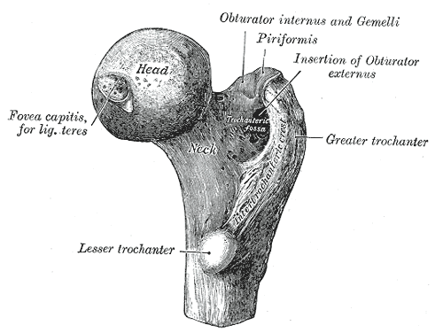
Neck
- Cylindrical strut, projects superomedially at 125 – 128 degree angle from the shaft (angle of inclination – decreases with age)
Greater trochanter
- Bony lateral projection from shaft, attaches hip abductors and external rotators
Lesser trochanter
- Posteromedial projection from shaft, inferior to the neck-shaft function
- Smaller than greater trochanter
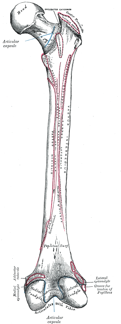
Intertrochanteric line
- A ridge of bone that runs in an inferomedial direction on the anterior surface of the femur, spanning between the two trochanters.
- After it passes the lesser trochanter on the posterior surface, it is known as the pectineal line
- It is the site of attachment for the iliofemoral ligament (the strongest ligament of the hip joint).
- It also serves as the anterior attachment of the hip joint capsule.
Intertrochanteric crest
- A ridge of bone that connects the two trochanters, located on the posterior surface of the femur. There is a rounded tubercle on its superior half called the quadrate tubercle; where quadratus femoris attaches.
Shaft
- Cylindrical
- Descends medially at 7 degrees from vertical with a mild anterior arch.
- Distally flares (cone-shaped) onto a cuboid base consisting of medial and lateral condyles, which articulate with the tibia to form the knee joint
- Linea aspera – Prominent, longitudinal bony ridge on the posterior surface, which diverges superiorly as pectineal line (medially) and gluteal tuberosity (laterally). Inferiorly it diverges as medial and lateral supracondylar lines
Medial and lateral condyles
- Rounded areas at the distal end of the femur, separated by the intercondylar fossa, form the articular surface with the menisci of the knee inferiorly and the patella anteriorly
- The medial condyle is larger, more narrow and further projected than its lateral counterpart, which accounts for the angle between the femur and the tibia.
Medial and lateral epicondyles
- Bony elevations on the non-articular surface, attaches the medial and lateral collateral ligaments
Articulations
- Hip joint – head with lunate surface of acetabulum
- Knee joint – condyles with menisci of tibia and patella
Muscular Attachments
- Greater trochanter: gluteus muscles (minimus, medius and maximus), external rotators (piriformis, obturator internus, gemellus superior and inferior), vastus lateralis
- Lesser trochanter: iliopsoas (iliacus, psoas major)
- Intertrochanteric crest: quadratus femoris
- Linea aspera: adductor muscles (longus, brevis, magnus), vastus lateralis, short head biceps femoris
- Lateral epicondyle: popliteus, plantaris
- Adductor tubercle: gastrocnemius (attaches behind)
Ligamentous Attachments
- Iliofemoral, pubofemoral, ischiofemoral ligaments
- Medial and lateral collateral ligaments of the knee
- Anterior and posterior cruciate ligament
- Oblique popliteal ligament
- Meniscofemoral ligament
Arterial Supply
Femoral head
- Medial and lateral femoral circumflex arteries (branch of deep femoral artery)
- Artery to ligamentum teres (branch of medical femoral circumflex artery)
- Genicular artery branches of ascending cervical
Knee
Development
- Ossifies in cartilage
- A center in the shaft appears in utero (8th week) and a center at distal femur appears at birth (end of 9th fetal month) with its presence a medicolegal evidence of maturity. The distal portion is the growing end with its epiphysis uniting with the shaft after 20 years.
A center appears in the femoral head at the age of 1, greater trochanter at 3, and lesser trochanter at 12 years. These fuse with the shaft at about 18 years of age.
Variants
- Cervical fossa (Allen’s fossa) – small depression usually located near the anterior superior margin of the femoral neck
- Poirer’s facet – slight bulging of the articular surface of the femoral head towards the anterior portion of the femoral neck
- Hypotrochanteric fossa – located in the superior posterior part of the femoral diaphysis between the gluteal ridge and the lateral margin
- Exostosis in trochanteric fossa – various degrees of bony spicules or exostosis
- Third trochanter – rounded tubercle at superior border of the glutal crest
References
Variants of the Femur
Michael Finnegan, M. A. Faust
https://scholarworks.umass.edu/cgi/viewcontent.cgi?article=1002&context=anthro_res_rpt14
Michael Finnegan, M. A. Faust
https://scholarworks.umass.edu/cgi/viewcontent.cgi?article=1002&context=anthro_res_rpt14