

Overview
The radius is a long bone in the forearm. It lies laterally and parallel to the ulnar. The long narrow medullary cavity is enclosed in a strong wall of compact bone, which is thickest along the interosseous border and thinnest at the extremities.
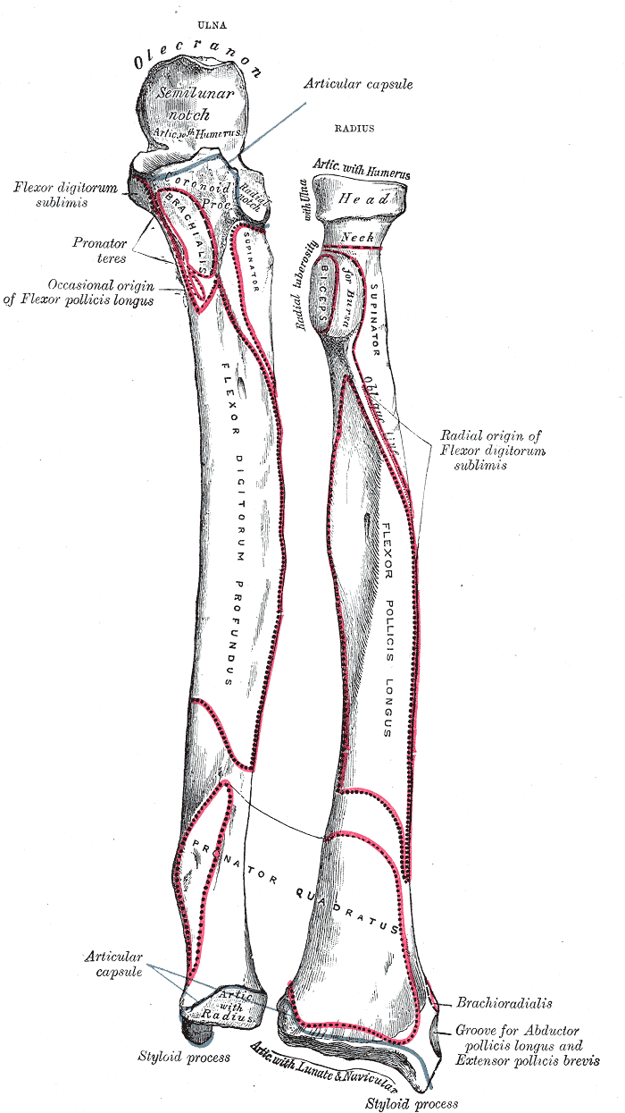
Surfaces
Volar (anterior) surface
- Rough and irregular
- Gives attachment to the volar radiocarpal ligmanet

Dorsal (posterior) surface
- Convex
- Gives attachment to the dorsal radiocarpal ligament and is marked by three grooves
- First groove – broad, shallow
- Second groove – deep but narrow
- Third groove – broad
Borders
Volar border
- Extends from the lower part of the tuberosity above to the anterior part of the base of the styloid process below
- Separates the volar surface from the lateral surface
- Its upper third is prominent, and from its oblique direction has received the name of the oblique line of the radius; it gives origin to the flexor digitorum superficialis muscle (also flexor digitorum sublimis) and flexor pollicis longus muscle; the surface above the line gives insertion to part of the supinator muscle.
- The middle third of the volar border is indistinct and rounded.
- The lower fourth is prominent, and gives insertion to the pronator quadratus muscle, and attachment to the dorsal carpal ligament; it ends in a small tubercle, into which the tendon of the brachioradialis muscle is inserted.
Dorsal border
- Begins above at the back of the neck, and ends below at the posterior part of the base of the styloid process
- Separates the posterior surface from the lateral surface.
- is indistinct above and below, but well-marked in the middle third of the bone
Interosseous border
- Begins above, at the back part of the tuberosity, and its upper part is rounded and indistinct; it becomes sharp and prominent as it descends, and at its lower part divides into two ridges which are continued to the anterior and posterior margins of the ulnar notch. To the posterior of the two ridges the lower part of the interosseous membrane is attached, while the triangular surface between the ridges gives insertion to part of the pronator quadratus muscle.
- Separates the volar surface from the dorsal surface, and gives attachment to the interosseous membrane. The connection between the two bones is actually a joint referred to as a syndesmosis joint.
Articulations
- Elbow Joint – partly formed by the articulation between the head of the radius, capitulum of the humerus
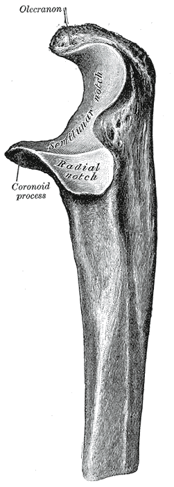
- Proximal Radioulnar Joint – articulation between the radial head and radial notch of the ulnar
- Wrist Joint – articulation between the distal end of the radius and carpal bones (scaphoid and lunate)

- Distal Radioulnar Joint – articulation between the ulnar notch and head of the ulnar
Structure
- Radial head – disc shaped structure with a concave articulating surface located proximally, articulates with the capitellum (or capitulum) of the humerus and the radial notch of the ulna. The head rotates within the annular ligament to produce supination and pronation of the forearm.
- Neck – narrowing between the head and radial tuberosity
- Radial (bicipital) tuberosity – oval-shaped convexity where the biceps brachii inserts
- Coronoid process
- Shaft – long wide column of bone that is convex laterally and enlarges towards the wrist
- Styloid process
- Ulnar notch – medial concavity upon the distal head which directly contacts the distal ulna
- Dorsal tubercle – protrudes on the posterior aspect of the distal head of the radius, seated between the grooves for the tendons of the extensor carpi radialis
Muscle Attachments
Proximal
- Biceps brachii attaches to the radial tuberosity.
- Supinator, flexor pollicis longus and the flexor digitorum superficialis attach to the upper third part of the shaft of the radius.
Mid shaft
- Extensor Pollicis Brevis muscle
- Abductor pollicis longus muscle
- Pronator teres
Distal
- Pronator quadratus muscle
- Tendon of the supinator longus