

Overview
The atlantooccipital joints (less commonly known as the C0-C1 joint) are a symmetrical condyloid synovial joint between the occipital bone (C0) and first cervical vertebra (atlas/C1), classified as a biaxial, pivot joint.
The upper cervical spine region is designed to allow for significant mobility, at the sacrifice of stability. This is why joint stability in the craniocervical region is maintained mainly by the fibrous capsules, ligaments, articular surfaces and surrounding muscles.
Articulations
Each joint is comprised of two concave articular surfaces on the superior aspect of the lateral mass of atlas which articulate with a convex surface on the occipital condyle. The joint is reinforced by fibrous capsules which support each joint. The atlanta facets are tilted medially.

Movements & Muscles
Movements
- The principal movement at the atlantooccipital joints is simultaenous flexion-extension (approximately 25°), permiting nodding of the head, and therefore functions mechanically as a single joint
- Limited axial rotation (5°)

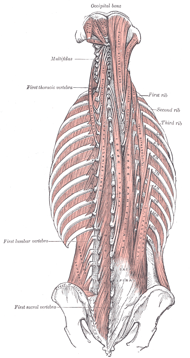
Muscles
| Flexion from the upright position | Trapezius, splenius capitis, longissimus capitis, semispinalis capitis, rectus capitis posterior major, rectus capitis posterior minor, obliquus capitis superior |
| Flexion from the supine position | Sternocleidomastoid, longus capitis, rectus capitis anterior muscles |
| Extension from the upright position | Sternocleidomastoid, longus capitis, rectus capitis anterior muscles |
| Extension from the prone position | Rectus capitis posterior major, rectus capitis posterior minor, obliquus capitis superior, semispinalis capitis, splenius capitis muscles, cervical part of trapezius |
| Lateral flexion | Rectus capitis lateralis, trapezius, splenius capitis, semispinalis capitis, sternocleidomastoid |
Attachments
Articular Capsule
- A loose, thin articular capsule, composed of fibrous tissue and lined with synovial membrane, which envelops each atlantooccipital joint
- It attaches to the margins of the articular facets
- Capsular thickenings can be noted on its posterior and lateral aspects.

Ligamentum nuchae
- A large, thick, strong midline ligament that is a superior and posterior extension of the supraspinous ligament, covering the spinous process tips from C7 to the inion of the occipital bone
- It limits hyperflexion of the neck.
Alar ligament
- The alar ligaments arise bilaterally from the upper posterior portion of the odontoid process of C2 and runs superolaterally to insert on the medial surface of the occipital condyles
- They stabilize the head during rotatory movements – limiting axial rotation and contralateral lateral flexion of the head.
- In conjunction with the transverse band of the cruciform ligament, they are the primary stabilizers of the atlantoaxial joint
Apical ligament
- A small ligament that joins the apex of the odontoid process of C2 to the anterior margin (basion) of the foramen magnum.
- It is the weak, fibrous remnant of the notochord and does not contribute significantly to stability.
- Anterior is the anterior atlanto-occipital membrane.
Lateral atlantooccipital ligament
The anterior and posterior atlanto-occpital membranes primarily connect the occipital bone with the atlas and are the principal ligaments of the atlantooccipital joint.

Anterior atlanto-occipital membrane
- Broad, dense fibrous structure which joins the upper border of the anterior arch of the atlas (C1) to the anterior inferior margin of the foramen magnum
- It is a continuation of the anterior longitudinal ligament and prevents excessive neck extension
- Laterally it blends with the joint capsule and medially is strengthened by a strong, rounded cord which connects the basilar occipital bone to the anterior atlantal tubercle.
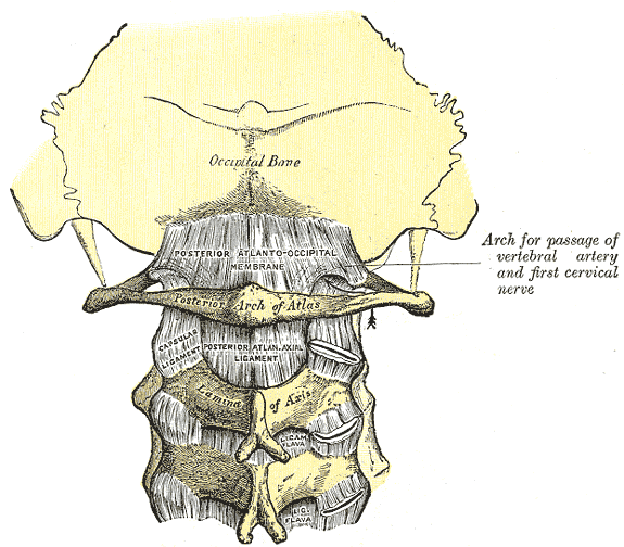
Posterior atlanto-occipital membrane
- A broad, thin fibrous membrane which attaches the upper border of the anterior surface of the posterior arch of the atlas (C1) to the posterior margin of the foramen magnum
- It is continuous with the posterior atlantoaxial membrane inferiorly (part of the ligamentum flavum) and ligamentum nuchae posteriorly and lies immediately posterior to the spinal dura.
- Posteriorly lie the suboccipital muscles.
- The atlantic (V3) portion of the vertebral artery runs anteriorly to pierce the membrane and dura to become the dural (V4) portion.
Tectorial membrane
- Continuation of the posterior longitudinal ligament from the dens to the anterior aspect of the foramen magnum.
Innervation
The joint capsule is innervated by the C1 nerve.
Arterial Supply
Anastomosis between the deep cervical, occipital and vertebral arteries.
Relations
- Posterior – vertebral artery
- Anterior – rectus capitis anterior
- Posteromedial – dorsal ramus of the first cervical nerve and rectus capitis posterior major
Anatomical Variants
- Variable degree of communication between the atlanto-occipital joint and the cavity between the dens (odontoid peg) and transverse ligament of the cruciform ligament
- Third occipital condyle (condylus tertius) which may articulate with the anterior arch of the atlas, or the dens
paracondylar process - Occipital condyle hypoplasia
- Division of the superior articular facet of the atlas
- Atlanto-occipital assimilation
- Arcuate foramen – foramen visualized by the atlantic portion of the vertebral artery as it courses through a calcified posterior atlanto-occipital membrane