

Overview
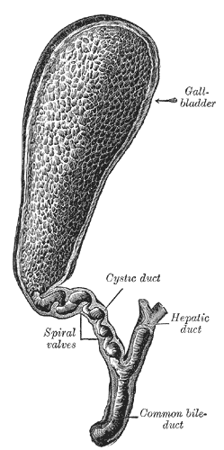
A distensible pear-shaped musclomebranous sac which stores and concentrates bile between meals.
Location
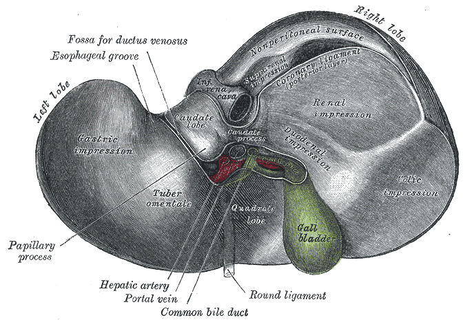
Gallbladder fossa on the visceral surface of the liver, to the right of the quadrate lobe. It extends from the porta hepatis to the anterior abdominal wall.
Structure
- Fundus – rounded blind-end, may project anterior from inferior border of liver
- Body – lies within the gallbladder fossa
- Neck – narrowing with mucosal fold forming spiral folds
- Cystic duct – continuous with neck, allowing bidirectional passage of bile into and from common hepatic duct
Blood supply
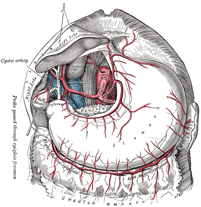
- Cystic artery – most commonly arises as a branch from right hepatic artery (derived from coeliac trunk, common hepatic artery then common hepatic artery)
Venous drainage
- Cystic vein – drains into the portal vein
Innervation
- Sympathetic: Coeliac plexus – formed in part by the greater and lesser splanchnic nerves of both sides, and fibres from the anterior and posterior vagal trunks
- Parasympathetic: Hepatic branches of the anterior vagal trunk
- Somatic: Right phrenic nerve – conveys sensory information
Lymphatic drainage
- Porta hepatis node and cystic node – drains into the coeliac group
Relations
- Superior: visceral surface of the liver
- Inferior: transverse colon near hepatic flexure, 2nd part of duodenum
- Medial: epiploic foramen, free margin of lesser omentum, 1st part of duodenum
- Lateral: right lobe of liver
- Anterior: anterior abdominal wall, visceral surface of liver
- Posteriorly: right kidney
Anatomical Variants
- Hartmann’s pouch – small outpouching between neck and cystic duct
- Septated gallbladder or Phrygian cap (septum between body and fundus)
- Ectopic gallbladder
- Accessory or aberrant drainage of cystic duct
- Cystic artery arising from left hepatic artery, hepatic artery proper or gastroduodenal artery
- Duplication/triplication/agenesis or hypoplasia