

Overview
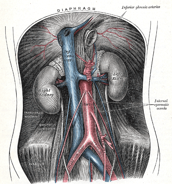
The kidneys are paired bean-shaped retroperitoneal organs located in the upper left and right abdominal quadrants, extending from T12 to L3 (right kidney may be relatively lower due to the presence of the liver). The superior poles of the kidneys are usually oriented slightly medially and posteriorly relative to the lower poles The dimensions are usually a length of 10 to 12 cm, a width of 5 to 7 cm, and a thickness of 3 to 5 cm. The kidney primarily functions to;
- Eliminate toxic metabolites through urine
- Regulate blood homeostasis and blood pressure
- Synthesise hormones (erythropoetin and renin)
- Convert precursor of vitamin D to active calcitriol
Major Structures
External Layers (superficial to deep)
- Renal capsule – tough fibrous capsule
- Perirenal fat – collection of extraperitoneal fat.
- Renal fascia (Gerota’s fascia or perirenal fascia) – encloses the kidneys and the suprarenal glands.
- Pararenal fat – mainly located on the posterolateral aspect of the kidney.

Parenchyma
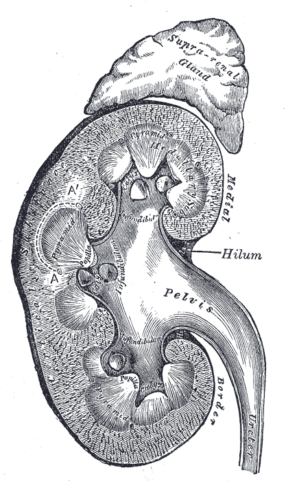
The renal parenchyma can be divided into two main areas – the outer cortex and inner medulla. The cortex extends into the medulla, dividing it into triangular shaped lobes known as renal pyramids. Between the renal pyramids are projections of cortex called renal columns.
Collecting System (macroanatomy)
- The renal papilla is the apex of the pyramids and empties urine into the minor calyx.
- Several minor calices merge to form a major calyx
- The major calices empty into the renal pelvis, a flattened and funnel-shaped structure.
- From the renal pelvis, urine drains into the ureter, which transports it to the bladder for storage.
Hilum
- The medial margin of each kidney is marked by a deep fissure, known as the renal hilum, at the level of L2. At the hilum, the ureter and renal vein exit the kidney and the renal artery enters. Hilar fat and lymphatic tissue with lymph nodes surround these structures.
- The hilar fat is contiguous with a fat-filled cavity called the renal sinus.
- The renal sinus collectively contains the renal pelvis and calyces and separates these structures from the renal medullary tissue.
- The most superior vessel is the renal vein, followed by the renal artery then the ureter.
- The anterior to posterior orientation also follows the same order: renal vein, renal artery and ureter.
Arterial Supply
The kidneys receive (renal blood flow) approximately 20 – 25% of cardiac output.
Renal Arteries
- Arise directly from lateral aspect of the abdominal aorta at the level of L1 – L2, immediately distal to the origin of the superior mesenteric artery.
- Due to the anatomical position of the abdominal aorta (slightly to the left of the midline), the right renal artery is longer, and crosses the inferior vena cava posteriorly.
- Enters the kidney via the renal hilum at the level of L2, forming an anterior and a posterior division, which carries 75% and 25% of the blood supply to the kidney, respectively.
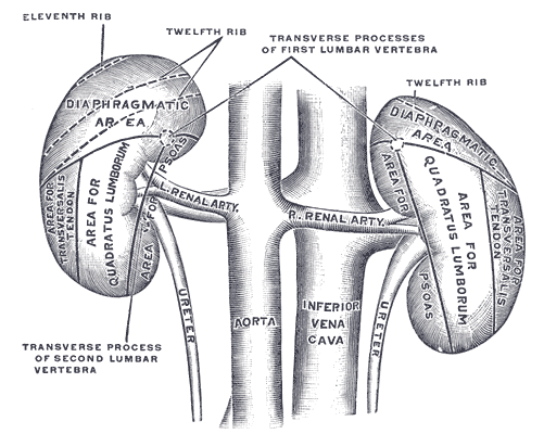
- The right renal artery courses inferiorly and passes posterior to the inferior vena cava and the right renal vein to reach the renal hilum. The left renal artery is much shorter and arises slightly more superior to the right main renal artery. The left renal artery courses more horizontally, posterior to the left renal vein to enter the renal hilum.
- The avascular plane of the kidney (line of Brodel) is a line along the lateral and slightly posterior border of the kidney, which delineates the segments of the kidney supplied by the anterior and posterior divisions.
Each renal artery gives off small branches (not visible on imaging) in its proximal course, prior to dividing into anterior and posterior divisions:
- Inferior adrenal artery – supplies the adrenal gland together with the superior suprarenal artery (arising from the inferior phrenic artery) and middle suprarenal artery (arising from the abdominal aorta)
- Ureteric artery
- Capsular artery
Segmental Arteries
- Five segmental arteries originate from the anterior and posterior divisions: apical, anterior superior, anterior inferior (middle), inferior and posterior segmental renal arteries
Lobar Arteries
- The segmental arteries give off lobar branches, usually one for each renal pyramid.
Interlobar Arteries
- Upon reaching the minor calyces, the lobar arteries further subdivide into interlobar arteries, situated either side every renal pyramid.
Arcuate Arteries
- Interlobar arteries undergo further division to form the arcuate arteries at the base of the pyramids.
Interlobular Arteries
- Arcuate arteries then enter the nephrons as the interlobular arteries, at 90 degrees to the arcuate arteries
- The interlobular arteries pass through the cortex, dividing one last time to form afferent arterioles.
Arterioles
- The afferent arterioles form a capillary network, the glomerulus, where filtration takes place.
- The capillaries come together to form the efferent arterioles.
- In the outer two-thirds of the renal cortex, the efferent arterioles form what is a known as a peritubular network, supplying the nephron tubules with oxygen and nutrients.
- The inner third of the cortex and the medulla are supplied by long, straight arteries called vasa recta.
Venous Drainage
- Drain by paired (left and right) renal veins, which leave the renal hilum anterior to the renal arteries, and empty directly into the inferior vena cava
- As the inferior vena cava lies slightly to the right, the left renal vein is longer and travels anterior to the abdominal aorta below the origin of the superior mesenteric artery
Innervation
- Renal plexus – fibres course along the renal arteries
- Input from the sympathetic nervous system triggers vasoconstriction in the kidney, thereby reducing renal blood
- Also receives input from the parasympathetic nervous system, by way of the renal branches of the vagus nerve
- Sensory input from the kidney travels to the T10-11 levels of the spinal cord and is sensed in the corresponding dermatome
Lymphatics
Lymph from the kidney drains into the lateral aortic lymph nodes, which are located at the origin of the renal arteries.
Relations
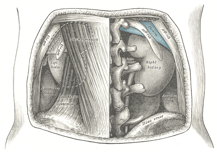
Left kidney
- Anterior: Suprarenal gland, connected to spleen (anterolaterally) by splenorenal ligaments, stomach, pancreas, jejunum, splenic flexure of the colon and descending colon
- Superiorly: Suprarenal glands (adrenal glands) separated by renal fascia
- Medial: Aorta, greater curvature of the stomach
Right kidney
- Anterior: Ascending colon, second part of the duodenum medially, liver – separated by the hepatorenal recess
Variant Anatomy
- Horsehoe kidney
- Pancake kidney
- Supernumerary kidney
- Duplicated collecting system (with single or two ureters)
- Accessory renal arteries (~30%) – usually arises just below the main renal artery. Rarely from coeliac trunk, superior mesenteric artery, or above the main renal artery.
- Aberrant renal arteries – enter via the renal capsule rather than the hilum
- Early-branching (or prehilar branching) (~10%)
- occurs within 1.5-2.0 cm of origin in the left renal artery or in the retrocaval segment of the right renal artery
- important to recognise in renal transplant for successful anastomoses
- Thoracic renal artery
- Aberrant origin of the renal artery:
- Bifurcation of the aorta or from the common iliac, internal iliac or inferior mesenteric artery.
- Retroarotic renal vein